THE AGING MUSCLE
Click image for closeup view Click pictures for new window with figure and legend, click again for high resolution image

1 General Description

C. elegans possess two types of muscle cells which are differentiated by the presence of a single or multiple sarcomeres, the basic element of the muscle contractile apparatus (See Hermaphrodite - Muscle Introduction for more details). Non-striated muscle cells, containing a single sarcomere, are found at several sites, including the muscles of the pharynx, the intestine, the anus, the gonad and the male tail. During aging, the pharynx muscles become disorganized and deformed (see Aging Pharynx). The second major C. elegans muscle group are the striated muscles, containing multiple sarcomeres, that compose the locomotory body wall muscles. Adults have 95 body wall muscles, making this the most abundant muscle type in C. elegans. Body wall muscle controls movement and locomotion, often in response to sensory stimuli.

The body wall muscles are arranged in 4 longitudinal bundles of staggered muscle cell pairs along the quadrants of the body cylinder. The muscle cell bundles are attached to the cuticle and hypodermis at multiple attachment points, allowing coupling of myofibril contraction with the organism’s movement. Intracellularly, each muscle cell contains the contractile myofilament lattice, which is an organized arrangement of parallel muscle filaments along the muscle cell boundaries. In muscle cells, the nucleus, mitochondria and other cytoplasmic components are located in the muscle cell body, or belly, which lacks contractile elements. At each cell boundary, cytoplasmic muscle arms extend towards the nerve cord and serve as the site for the neuromuscular junction (See Hermaphrodite - Somatic Muscle; AMusFIG 1).

AMusFIG 1 C. elegans body wall muscles
AMusFIG 1: C. elegans body wall muscles. A. The organization of somatic muscles in the adult C. elegans hermaphrodite, dorsal oblique view, visualized using epifluorescence in a transgenic animal expressing the unc27::GFP reporter gene. The body wall muscles are organized in four quadrants (only the dorsal quadrants are visible here) with two rows of cells in each. The quadrants are placed subdorsally and subventrally. The two dorsal quadrants flank the dorsal hypodermal ridge and the dorsal cord, and the ventral quadrants flank the ventral hypodermal ridge and the ventral nerve cord. Anteriorly, the spindle-shaped cells in each quadrant are arranged almost in pairs, whereas more posteriorly the cells are organized in an alternating fashion. (Strain source: L. Jia and S.W. Emmons.) Bar, 50 m. B. Low-power TEM showing the relationship of the muscle quadrants to the hypodermis and internal organs, transverse section. Note the somewhat flattened profile of a normal muscle nucleus. Bar, 1 m. (Image source: [Hall] N501-N354.) C. Three-dimensional rendering of myofilament lattice as well as structure of the sarcomere. (Yellow lines) Thick filaments; (black lines) thin filaments; (black dots) dense body (DB); (brown dots) M lines. D. TEM cross section of the contractile apparatus. The filaments of the lattice are oriented longitudinally in rows that are perpendicular to the surface. Dense bodies (DBs) anchor the thin (actin) filaments, whereas M line homologs anchor the thick (myosin) filaments. The membranous sacs of sarcoplasmic reticulum (SR) align around the dense body and are also present under the thick and thin filament bands along the muscle membrane. A single unit of myofilament lattice between two DBs is called a sarcomere and contains one A band in the middle and two juxtaposing half I bands. In C. elegans, each adult body wall sarcomere is about 1 µm wide. Bar, 0.5 m. (Image source: [Hall] N501C.)

2 Structural Changes in Body Muscle During Aging

C. elegans body muscle cells can exhibit significant structural deterioration during aging. In the sarcomeric region, this deterioration is stochastic and varies among individual cells within one animal. Sarcomeres from older adults may appear thinner and disorganized (Herndon et al., 2002; Garigan et al., 2002; Glenn et al 2004; Bansal et al., 2015; Herndon et al., 2017; Dhondt et al., 2021) (AMusFIG 2, AMusFIG 3 & AMusFIG 4). Myofilament bundles persist although they may appear to contain fewer myosin thick filaments. There may be a loss of parallel fiber arrangement, with fibers appearing broken in rare cases (AMusFIG 2E). However, these appear to be localized changes and aged sarcomeres appear more or less intact in cross-sectional views (AMusFIG 3, AMusFIG 4 & AMusFIG 5) and despite this cellular disorganization, aging muscle cells appear to maintain the structures that connect muscle to the cuticle (see Aging Hypodermis) (Herndon et al., 2002).

AMusFIG 2: Body wall sarcomeres in aging agnimals
AMusFIG 2: Body wall sarcomeres in aging animals. A&B. Muscle cell aging visualized using a MYO-3::GFP translational fusion highlighting the myosin heavy chain A in body wall sarcomeres from a young day 1 adult (A) and an aged day 15 adult, with evidence of sarcomere disorganization (B). (Image source: Herndon et al., 2002.) C-E. Electron micrographs showing longitudinal organization of sarcomeres. C. Muscle fibers in a young adult are evenly organized and are closely opposed to the contents of the muscle belly (black arrow). (Image source: N533 [Hall] 4235.) D. Muscle fibers in older adults (15 days) are less cohesive and the muscle belly is grossly shrunken with few organelles seen (black arrow), so that the muscle cell plasma membrane (where it opposes the pseudocoelom) often seems collapsed against the sarcomere. (Image source: N815 [Hall] G713, Class B.) E. In rare cases myosin filaments are frayed and bent (white arrow). (Image source: N803 [Hall] E769, Class C.)

Similar changes in muscle fiber structure are seen in aging animals throughout the body from head (AMusFIG 3), midbody (AMusFIG 4) and tail (AMusFIG 5) regions with a greater deterioration of structure and organization of muscle fibers correlating with increased age and behavioral class of the animal (Herndon et al., 2002). More substantial deterioration first occurs in the muscle belly, the non-contractile portion of the muscle cell. In young adults, the muscle belly is replete with subcellular components, including many mitochondria (AMusFIG 3 & AMusFIG 4). In older adults, the muscle belly appears shriveled and with a loss of cytoplasmic components (AMusFIG 3B, AMusFIG 4B & AMusFIG 5C&D).

Nuclei may appear misshapen in aged adults, with enlargement and fragmentation of the nucleolus observed (Herndon et al., 2002; Haithcock et al., 2005) (AMusFIG 6). Some studies have found that smaller nucleolar size is a hallmark of metabolic health and can be predictive of longevity both in C. elegans and in other taxa (Tiku et al., 2017). Nuclear lamina morphology is also altered during aging (Haithcock et al., 2005). While nuclear structure phenotypes were variable within muscle cells of an individual animal, locomotory decline generally correlated with more severe structural declines in the body muscle nuclei and nuclear lamina (Herndon et al,. 2002; Haithcock et al., 2005).


AMusFIG 3: Muscles in the head of young and old adult animials.

AMusFIG 3: Muscles in the head of young and old C. elegans. Illustrations show (arrow) approixmate locations for the transverse micrographs through the head of a young adult (A) and an older, day-15 adult (B). Enlargements of boxed regions (right panels) show detail of body muscle cells. B. Sarcomeres (S) are reduced in the aged muscle, muscle cell body (MB) is shrunken and empty of subcellular components, and mitochondrial (M) density may be reduced. A prominent nucleus, somewhat distorted in shape (compare with AMusFIG 1B) and containing an enlarged nucleolus, lies beneath the sarcomeres in the muscle belly while the myofilament lattice is smaller and disorganized, including a dramatic decline in myosin filaments per sarcomere. Green shading indicates muscles; medial darker green region is the pharynx; lighter green indicates the four body muscle quadrants at the body periphery. Both the pharyngeal and body wall muscles exhibit significant deterioration in the aged animal. (Image source: A. N2T [Hall] 374 - Note that N2T was not fixed as well as the other examples and shows rather light staining of the muscle belly cytoplasm compared to other images in this chapter. B. Left panel N813 [Hall] G515 Bar, 5 µm. Right panel N813 [Hall] G516. Bar, 1µm. Class C.)

AMusFIG 4: Muscles in the midbody region of young and old adult animals
AMusFIG 4: Muscles in the midbody region of young and old C. elegans. A. Illustration of young adult with arrow indicating approximate location in the midbody region shown in micrograph below. Left panel shows low resolution view of image section in transverse view. Box indicates region enlarged in right panel. Bar, 5 m. Right panel shows higher magnification view of body muscle. Green outline, body muscle cell; lavender, intestine; blue, germline; purple, somatic gonad. S, sarcomere; M, mitochondrion; MB, muscle belly. Bar, 1 m. (Image source: N506 [Hall] M676.) B. Illustration of older adult with arrow indicating approximate positions of micrographs shown in panels i, ii & iii below. Lower panels show higher-magnification views of midbody muscle cells from older day 15 adults in longitudinal view (i) or transverse view (ii & iii). i. The muscle belly is shrunken though a nucleus containing a large nucleolus (similar to what is seen in AMusFIG 3B) is found close to the muscle fibers. Close to the muscle cell is a large volume of dark yolk material that has accumulated. ii. While a more significant muscle belly is featured in this animal and mitochondria are present, the contents appear disorganized with wispy pieces of membrane. iii. Muscle belly is shrunken with few organelles or electron dense components other than a few small mitochondria. Note that in all cases the muscle fibers retain their attachments to the hypodermis, but the cuticle is vastly thickened. S, sarcomere; M, mitochondrion; MB, muscle belly; N, nucleus. (Image sources: B. N803 [Hall] E797; C. N810 [Hall] E345; D. N810 [Hall] M783. Bars, 1 µm.)

AMusFIG 5: Muscles in the rectal region of young and old adult <em>C. elegans</em>
AMusFIG 5: Muscles in the rectal region of young and old C. elegans. A. Illustration showing anatomy in the rectal area, containing the hindgut. For more information, refer to Alimentary System - Rectum. B. Illustration of young adult with arrow indicating approximate location in the tail region shown in micrograph below showing a transverse view of the rectum. (Image source: B140C [Hall] T565.) C & D. Illustration of old adult with arrow indicating approximate location in the tail region shown in micrographs below. Micrographs are transverse views of rectal area from two older (day 15) adult animals. While the anal depressor muscle is still present and appears to retain its attachments, the muscle cells appear withered and disorganized and contain less electron dense material in C and D, but especially pronounced in D from a class B animal. Darker green, body wall muscle cells; lighter green, anal muscle cells. Scale bars, 5 µm. (Image sources: C. N826 [D. Hall] G5802, Class A; D. N801 [Hall] E556, Class B.)
AMusFIG 6: Body wall muscle cell nuclei in aging animals.
AMusFIG 6: Body wall muscle cell nuclei in aging C. elegans. Muscle nuclei visualized with MY0-3::GFP nuclear-localized reporter expressed in body muscle cells. Images of nuclear GFP in bodywall muscle cells from several individual young adults (A), middle-aged 7-day old adults (B) and older 15-day adults (C). In comparison to young adults, where chromatin fills the entire volume except for the nucleolus, the pattern of nuclear GFP in bodywall muscles becomes progressively fragmented and dispersed as animals age. Interestingly, individual nuclei from cells within one animal may develop this nuclear fragmentation at different rates. However, the progression of nuclear GFP fragmentation was correlated with the animals locomotory class (Herndon et al., 2002).

Mitochondrial dysfunction is hypothesized to be an important contributor to sarcopenia (Alway et al., 2017; Coen et al., 2019; Garcia-Casas et al., 2021; Sudevan et al., 2019). Muscle mitochondrial appear tubular in highly interconnected networks which undergo a progressive loss of connectivity as well as a reduction in mitochondrial content with increasing age (Weir et al., 2017). This structural change and appearance of fragmented mitochondria precedes sarcomeric decline (Regmi et al., 2014, Mergoud dit Lamarche et al., 2018) and correlates with lifespan and loss of movement (Weir et al., 2017; Gaffney et al., 2018; Mallick et al., 2020). Interventions that affect mitochondrial function also could preserve muscle cell structure and contractile function in older adults (Wang et al., 2019; Garcia-Casas et al., 2021).

Globally, aged cells demonstrate greater sensitivity to protein aggregation. Autophagy, the process which allows cells to remove dysfunctional cellular components, is reduced in muscle cells from older adults (Chang and Hansen, 2018). Proteasomal activity which removes aggregated proteins also appears to be reduced in muscle cells from older adults (Kern et al., 2010; Koyuncu et al., 2021). The combination of sarcomeric damage accumulated over a lifespan with aging-related declines in removal of damaged cell components may combinatorially drive losses of locomotory ability during aging in C. elegans.


3 Functional Decline of Body Muscle During Aging

During aging, C. elegans adults exhibit characteristic behavioral declines attributable to losses in body wall cell function which resemble aging-related decline of muscle strength and coordination in humans (AMusVID 1, see also Aging Introduction, AIntroVID 2, AIntroVID 3). C. elegans adults have progressive declines in the ability to perform normal sinusoidal movement as they age, eventually becoming unable to move forward or in reverse for any distance. At advanced ages, locomotory ability is lost and animals are left only with the ability to contract their body in response to physical stimuli (Croll et al., 1977; Bolanowski et al., 1981; Johnson, 1987; Duhon and Johnson, 1995; Herndon et al., 2002; Glenn et al., 2004; Huang et al., 2004; see AIntroVID 4). Locomotory decline is correlated with shorter chronological lifespan, and mirrors sarcomere disruption and muscle cell deterioration (Herndon et al., 2002; Johnston et al., 2008; Dhondt et al., 2021). Neuromuscular signaling also changes during aging, and may contribute to loss of locomotory behavior (Glenn et al., 2004; Liu et al., 2013). Interestingly, this pattern of structural and cellular muscle decline during aging is affected by the types of activity the animal experiences during its lifetime with increased muscle health associated with higher activity rates in C. elegans (Chuang et al., 2016; Laranjeiroa et al., 2017; Hartman et al., 2018; Laranjeiroa et al., 2019).


AMusVID 1. C. elegans movement declines during aging. Videos of swimming wildtype C. elegans (A) young adults (day 4) (B) middle-aged adults (day 11) and (C) old adults (day 15). The swimming movement is termed “thrashing” and can be manually counted or computationally analyzed in detail. Thrashing rates decline as the animals age. (Video Source: C.I. Ventoso and M. Driscoll, Rutgers University; Restif et al., 2014; Ibáñez-Ventoso et al., 2016)


4 References

Alway, S.E., Mohamed, J.S. and Myers, M.J. 2017. Mitochondria initiate and regulate sarcopenia. Exerc. Sport. Sci. Rev. 45: 58-69. doi: 10.1249/JES.0000000000000101

Bansal, A., Zhu, L.J., Yen, K. and Tissenbaum, H.A. 2015. Uncoupling lifespan and healthspan in Caenorhabditis elegans longevity mutants. PNAS. 112: E277-E286. doi: 10.1073/pnas.1412192112

Bolanowski, M.A., Russell, R.L. and Jacobson, L.A. 1981. Quantitative measures of aging in the nematode Caenorhabditis elegans. I. Population and longitudinal studies of two behavioral parameters. Mech. Ageing Dev. 15: 279-95. doi: 10.1016/0047-6374(81)90136-6 Article-pdf

Chang, J.T. and Hansen, M. 2018. Age-associated and tissue-specific decline in autophagic activity in the nematode C. elegans. Autophagy 14: 1276-1277. doi: 10.1080/15548627.2018.1445914

Chuang, H.S., Kuo, W.J., Lee, C.L., Chu, I.H. and Chen, C.S. 2016. Exercise in an electrotactic flow chamber ameliorates age-related degeneration in Caenorhabditis elegans. Sci. Rep. 6: 28064. doi: 10.1038/srep28064

Coen, P.M., Musci, R.V., Hinkley, J.M. and Miller, B.F. 2019. Mitochondria as a target for mitigating sarcopenia. Front. Physiol. 9: 1883. doi: 10.3389/fphys.2018.01883

Croll, N.A., Smith, J.M. and Zuckerman, B.M. 1977. The aging process of the nematode Caenorhabditis elegans in bacterial and axenic culture. Exp. Aging Res. 3: 175–189. doi: 10.1080/03610737708257101 Article-pdf

Dhondt, I., Verschuuren, C., Zecic, A., Loier, T., Braeckman, B.P. and De Vos, W.H. 2021. Prediction of biological age by morphological staining of sarcopenia in Caenorhabditis elegans. Dis. Model Mech. 14: dmm049169. doi: 10.1242/dmm.049169

Duhon, S.A. and Johnson, T.E. 1995. Movement as an index of vitality: comparing wild type and the age-1 mutant of Caenorhabditis elegans. J. Gerontol. Biol. Sci. 50: B254-B261. doi: 10.1093/gerona/50a.5.b254

Gaffney, C.J., Pollard, A., Barratt, T.F., Constantin-Teodosiu, D., Greenhaff, P.L. and Szewczyk, N.J. 2018. Greater loss of mitochondrial function with ageing is associated with earlier onset of sarcopenia in C. elegans. Aging 10: 3382-3396. doi: 10.18632/aging.101654

Garcia-Casas, P., Alvarez-Illera, P., Gomez-Orte, E., Cabello, J., Fonteriz, R.I., Montero, M. and Alvarez, J. 2021. The mitochondrial Na+/Ca2+ exchanger inhibitor CGP37157 preserves muscle structure and function to increase lifespan and healthspan in Caenorhabditis elegans. Front. Pharmacol. 12: 695687. doi: 10.3389/fphar.2021.695687

Garigan, D., Hsu, A.L., Fraser, A.G., Kamath, R.S., Ahringer, J. and Kenyon, C. 2002. Genetic analysis of tissue aging in Caenorhabditis elegans: a role for heat-shock factor and bacterial proliferation. Genetics 161:1101–1112. doi: 10.1093/genetics/161.3.1101 Article-pdf

Glenn, C.F., Chow, D.K., David, L., Cooke, C.A., Gami, M.S., Iser, W.B., Hanselman, K.B., Goldberg, I.G. and Wolkow, C.A. 2004. Behavioral deficits during early stages of aging in Caenorhabditis elegans result from locomotory deficits possibly linked to muscle frailty. J. Gerontol. 59A: 1251-60. doi: 10.1093/gerona/59.12.1251 Article-pdf

Haithcock, E., Dayani, Y., Neufeld, E., Zahand, A.J., Feinstein, N., Mattout, A., Gruenbaum, Y. and Liu, J. 2005. Age-related changes of nuclear architecture in Caenorhabditis elegans. PNAS 102: 16690-16695. doi: 10.1073/pnas.0506955102

Hartman, J.H., Smith, L.L., Gordon, K.L., Laranjeiro, R., Driscoll, M., Sherwood, D.R. and Meyer, J.N.  2018. Swimming exercise and transient food deprivation in Caenorhabditis elegans promote mitochondrial maintenance and protect against chemical-induced mitotoxicity. Sci. Rep. 8: 8359. doi: 10.1038/s41598-018-26552-9

Herndon, L.A., Schmeissner, P.J., Dudaronek, J.M., Brown, P.A., Listner, K.M., Sakano, Y., Paupard, M.C., Hall, D.H. and Driscoll, M. 2002. Stochastic and genetic factors influence tissue-specific decline in ageing C. elegans. Nature 419: 808-814. doi: 10.1038/nature01135 Article-pdf

Herndon, L.A., Wolkow, C.A., Driscoll, M. and Hall, D.H. 2017. Effects of ageing on the basic biology and anatomy of C. elegans. In Ageing: lessions from C. elegans. (ed Olsen, A. and Gill, M.). Chapter 2. pp. 9-39. Springer International, Switzerland. doi: 10.1007/978-3-319-44703-2_2 Article-pdf

Huang, C., Xiong, C. and Kornfeld, K. 2004. Measurements of age-related changes of physiological processes that predict lifespan of Caenorhabditis elegans. Proc. Natl. Acad. Sci. U S A. 101:8084–8089. doi: 10.1073/pnas.0400848101 Article-pdf

Ibáñez-Ventoso, C., Herrera, C., Chen, E., Motto, D. and Driscoll, M. 2016. Automated analysis of C. elegans swim behavior using CeleST software. J. Vis. Exp. 118: 54359. doi: 10.3791/54359

Johnson, T.E. 1987. Aging can be genetically dissected into component processes using long-lived lines of Caenorhabditis elegans. Proc. Natl. Acad. Sci. USA 84: 3777-81. doi: 10.1073/pnas.84.11.3777

Johnston, J., Iser, W.B., Chow, D.K, Goldberg, I.G. and Wolkow, C.A. 2008. Quantitative image analysis reveals distinct structural transitions during aging in Caenorhabditis elegans tissues. PLoS ONE. 3: e2821. doi: 10.1371/journal.pone.0002821 Article-pdf

Kern, A., Ackermann, B., Clement, A.M., Duerk, H. and Behl, C. 2010. HSF-1-controlled and age-associated chaperone capacity in neurons and muscle cells of C. elegans. PLoS ONE. 5: e8568. doi: 10.1371/journal.pone.0008568

Koyuncu, S., Loureiro, R., Lee, H.J., Wagle, P., Krueger, M. and Vilchez, D. 2021. Rewiring of the ubiquitinated proteome determines ageing in C. elegans. Nature. 596: 285-290. doi: 10.1038/s41586-021-03781-z

Laranjeiro, R., Harinath, G., Burke, D., Braeckman, B.P. and Driscoll, M. 2017. Single swim sessions in C. elegans induce key features of mammalian exercise. BMC Biol. 15: 30. doi: 10.1186/s12915-017-0368-4

Laranjeiro, R., Harinath, G., Hewitt, J.E., Hartman, J.H., Royal, M.A., Meyer, J.N., Vanapalli, S.A. and Driscoll, M. 2019. Swim exercise in Caenorhabditis elegans extends neuromuscular and gut healthspan, enhances learning ability, and protects against neurodegeneration. Proc. Natl. Acad. Sci. USA. 116: 23829-23839. doi: 10.1073/pnas.1909210116

Liu, J., Zhang, B., Lei, H., Feng, Z., Liu, J., Hsu, A.L and Xu, X.Z. 2013. Functional aging in the nervous system contributes to age-dependent motor activity decline in C. elegans. Cell Metab. 18: 392-402. doi: 10.1016/j.cmet.2013.08.007

Mallick, A., Ranawade, A., van den Berg, W. and Gupta, B.P. 2020. Axin-mediated regulation of lifespan and muscle health in C. elegans requires AMPK-FOXO signaling. iScience. 23: 101843. doi: 10.1016/j.isci.2020.101843

Mergoud dit Lamarche, A., Molin, L., Pierson, L., Mariol, M.-C., Bessereau, J.-L., Gieseler, K. and Solari, F. 2018. UNC-120/SRF independently controls muscle aging and lifespan in Caenorhabditis elegans. Aging Cell 17: 12713. doi: 10.1111/acel.12713

Regmi, S.G., Rolland, S.G. and Conradt, B. 2014. Age-dependent changes in mitochondrial morphology and volume are not predictors of lifespan. Aging. 6: 118-130. doi: 10.18632/aging.100639

Restif, C., Ibáñez-Ventoso, C., Vora, M.M., Guo, S., Metaxas, D. and Driscoll, M. 2014. CeleST: computer vision software for quantitative analysis of C. elegans swim behavior reveals novel features of locomotion. PLoS Comput. Biol. 10: e1003702. doi: 10.1371/journal.pcbi.1003702

Sudevan, S., Takiura, M., Kubota, Y., Higashitani, N., Cooke, M., Ellwood, R.A., Etheridge, T., Szewczyk, N.J. and Higashitani, A. 2019. Mitochondrial dysfunction causes Ca2+ overload and ECM degradation-mediated muscle damage in C. elegans. FASEB J. 33: 9540-9550. doi: 10.1096/fj.201802298R

Tiku, V., Jain, C., Raz, Y., Nakamura, S., Heestand, B., Liu, W., Spth, M., Suchiman, H.E.D., Mller, R.U., Slagboom, P.E., Partridge, L. and Antebi, A. 2017. Small nucleoli are a cellular hallmark of longevity. Nat Commun. 8: 16083. doi: 10.1038/ncomms16083

Wang, H., Webster, P., Chen, L. and Fisher, A.L. 2019. Cell-autonomous and non-autonomous roles of daf-16 in muscle function and mitochondrial capacity in aging C. elegans. Aging. 11: 2295-2311. doi: 10.18632/aging.101914

Weir, H.J., Yao, P., Huynh. F.K., Escoubas, C.C., Goncalves, R.L., Burkewitz, K., Laboy, R., Hirschey, M.D. and Mair, W.B. 2017. Dietary restriction and AMPK increase lifespan via mitochondrial network and peroxisome remodeling. Cell Metab. 26: 884-96. doi: 10.1016/j.cmet.2017.09.024


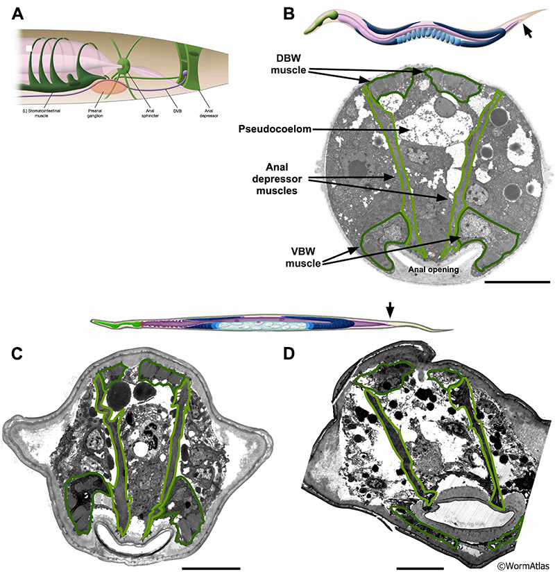
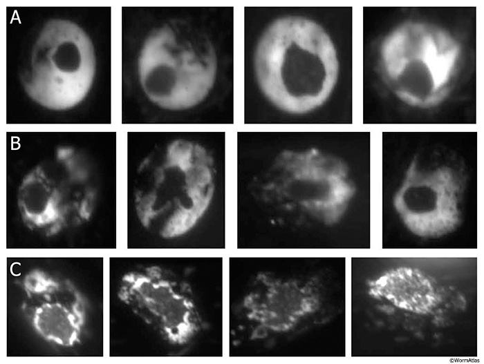
* Description of Behavioral Classes (A, B, C) as described in Herndon et al., 2002

To characterize aging phenotypes, age-synchronized individual worms were scored both for spontaneous movement and for response to prodding with a wire over the course of their lifespan. Three distinct classes representing behavioral phenotypes were established. Animals that move constantly and make sinusoidal tracks were designated as class A. Class B animals mainly move when prodded. When they move it is with uncoordinated motion, leaving non-sinusoidal tracks. Class C animals do not move forward or backward, even upon prodding, but do show head and/or tail movement and twitch in response to touch.  All animals begin adulthood in class A. Class B animals appear around days 6-7 of adulthood  and class C around day 9-10 (at 20oC). At later ages, animals representing all classes can be found within the same population and it was found that the behavioral class type was the better predictor of life expectancy than chronological age (Herndon et al., 2002). Due to the stochastic nature of the aging process in an individual nematode, these classifications only reflect ongoing changes in nerve and muscle, while other tissues can show very different age-related effects within one behavioral class, declining faster or remaining healthy much longer.


This chapter should be cited as: Herndon, L.A., Wolkow, C.A. and Hall, D.H. 2023. The aging muscle. In WormAtlas. doi:10.3908/wormatlas

Edited for the web by Laura A. Herndon. Last revision: January 20, 2023
image
 
image