|
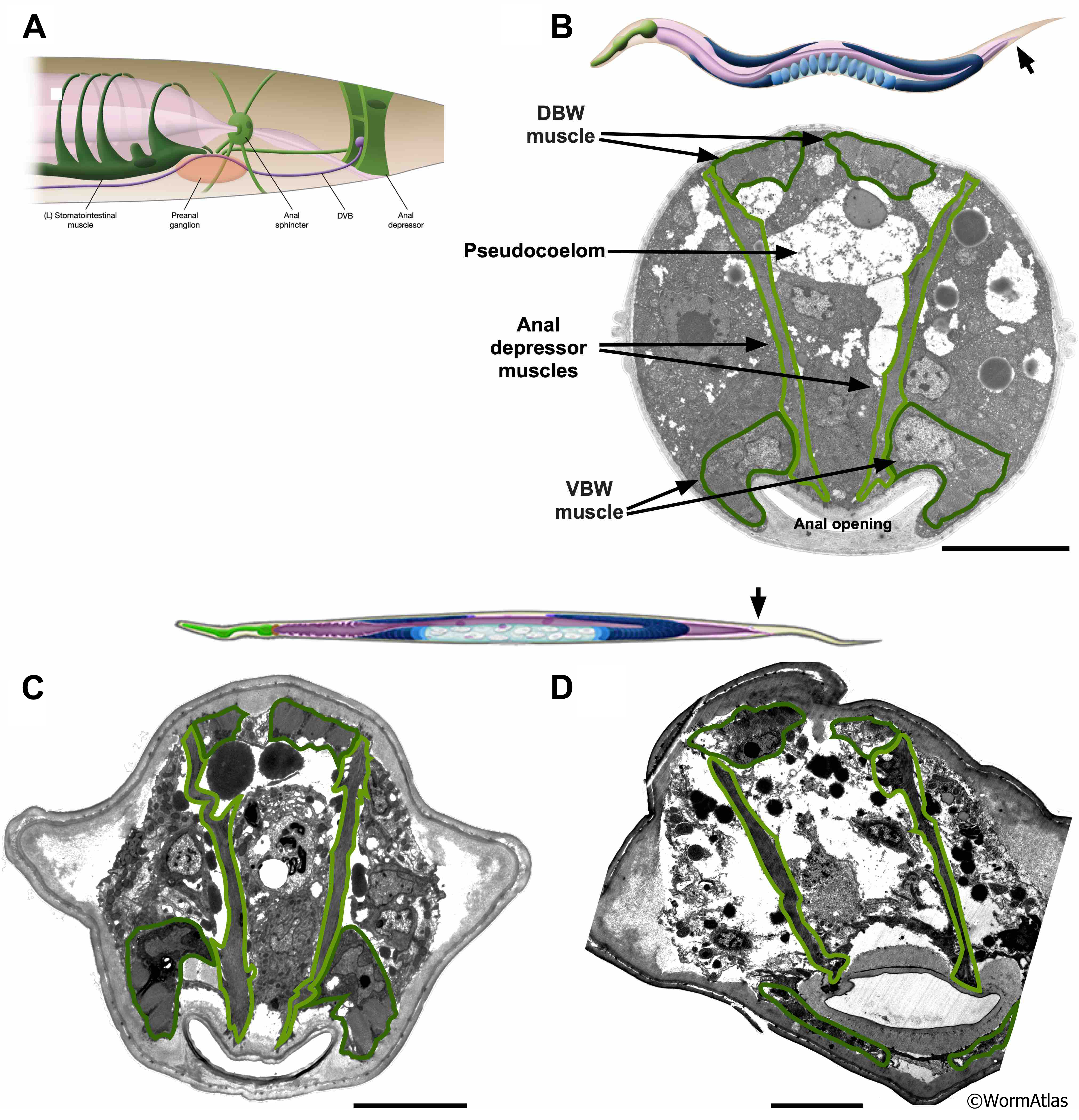
AMusFIG 5: Muscles in the rectal region of young and old adult C. elegans.
A. Illustration showing anatomy in the rectal area, containing the hindgut. For more information, refer to Alimentary System - Rectum.
B. Illustration of young adult with arrow indicating approximate location in the tail region shown in micrograph below showing a transverse view of the rectum. (Image source: B140C [Hall] T565.)
C & D. Illustration of old adult with arrow indicating approximate location in the tail region shown in micrographs below. Micrographs are transverse views of rectal area from two older (day 15) adult animals. While the anal depressor muscle is still present and appears to retain its attachments, the muscle cells appear withered and disorganized and contain less electron dense material in C and D, but especially pronounced in D from a class B animal.
Darker green, body wall muscle cells; lighter green, anal muscle cells. Scale bars, 5 µm. (Image sources: C. N826 [D. Hall] G5802, Class A; D. N801 [Hall] E556, Class B.)
Click on picture for full resolution image.
|