|
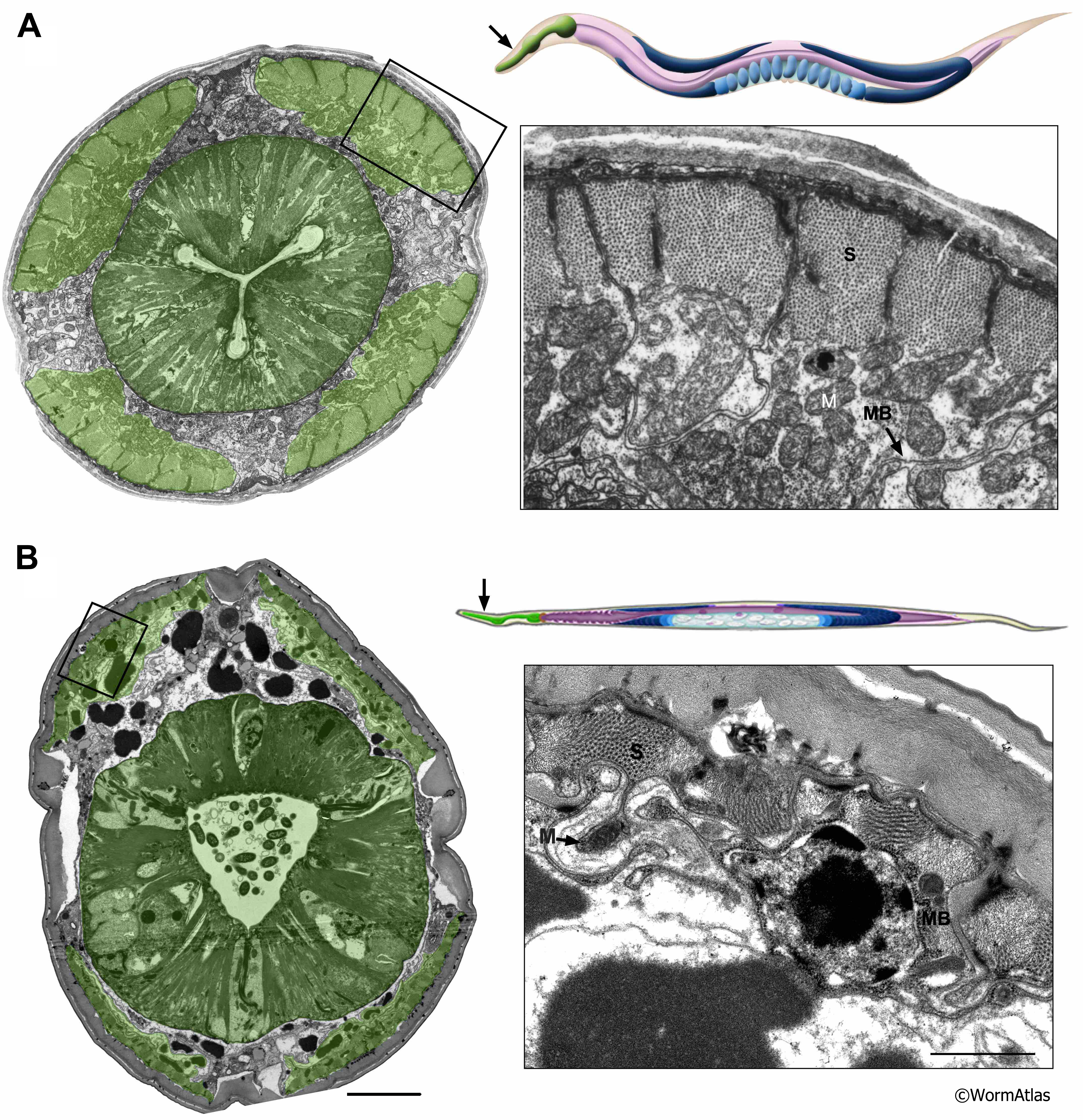
AMusFIG 3: Muscles in the head of young and old C. elegans.
Illustrations show (arrow) approixmate locations for the transverse micrographs through the head of a young adult (A) and an older, day-15 adult (B). Both the pharyngeal and body wall muscles exhibit significant deterioration in the aged animal. Enlargements of boxed regions (right panels) show detail of body muscle cells.
B. Sarcomeres (S) are reduced in the aged muscle, muscle cell body (MB) is shrunken and empty of subcellular components, and mitochondrial (M) density may be reduced. A prominent nucleus, somewhat distorted in shape (compare with AMusFIG 1B) and containing an enlarged nucleolus, lies beneath the sarcomeres in the muscle belly while the myofilament lattice is smaller and disorganized, including a dramatic decline in myosin filaments per sarcomere.
Green shading indicates muscles; medial darker green region is the pharynx; lighter green indicates the four body muscle quadrants at the body periphery.
(Image source: A. N2T [Hall] 374 - Note that N2T was not fixed as well as the other examples and shows rather light staining of the muscle belly cytoplasm compared to other images in this chapter. B. Left panel N813 [Hall] G515 Bar, 5 µm. Right panel N813 [Hall] G516. Bar, 1µm. Class C.)
Click on picture for full resolution image.
|