|
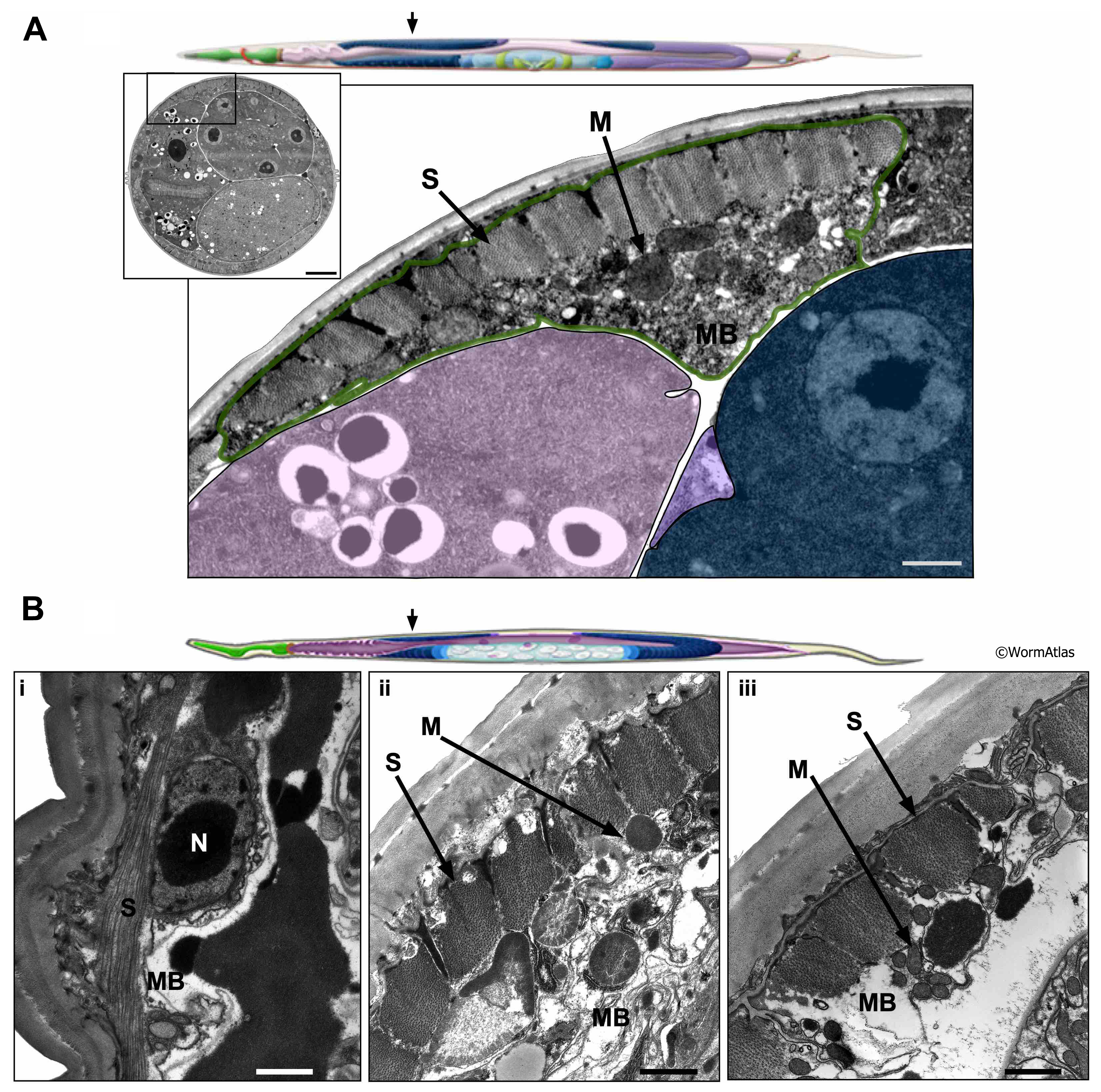
AMusFIG 4: Muscles in the head of young and old C. elegans.
A. Illustration of young adult with arrow indicating approximate location in the midbody region shown in micrograph below. Left panel shows low resolution view of image section in transverse view. Box indicates region enlarged in right panel. Bar, 5 µm. Right panel shows higher magnification view of body muscle.
Green outline, body muscle cell; lavender, intestine; blue, germline; purple, somatic gonad. S, sarcomere; M, mitochondrion; MB, muscle belly. Bar, 1 µm. (Image source: N506 [Hall] M676.)
B. Illustration of older adult with arrow indicating approximate positions of micrographs shown in panels i, ii & iii below. Lower panels show higher-magnification views of midbody muscle cells from older day 15 adults in longitudinal view (i) or transverse view (ii & iii). i. The muscle belly is shrunken though a nucleus containing a large nucleolus (similar to what is seen in AMusFIG 3B) is found close to the muscle fibers. Close to the muscle cell is a large volume of dark yolk material that has accumulated. ii. While a more significant muscle belly is featured in this animal and mitochondria are present, the contents appear disorganized with wispy pieces of membrane. iii. Muscle belly is shrunken with few organelles or electron dense components other than a few small mitochondria. Note that in all cases the muscle fibers retain their attachments to the hypodermis, but the cuticle is vastly thickened.
S, sarcomere; M, mitochondrion; MB, muscle belly; N, nucleus. (Image sources: B. N803 [Hall] E797; C. N810 [Hall] E345; D. N810 [Hall] M783. Bars, 1 µm.)
Click on picture for full resolution image.
|