|
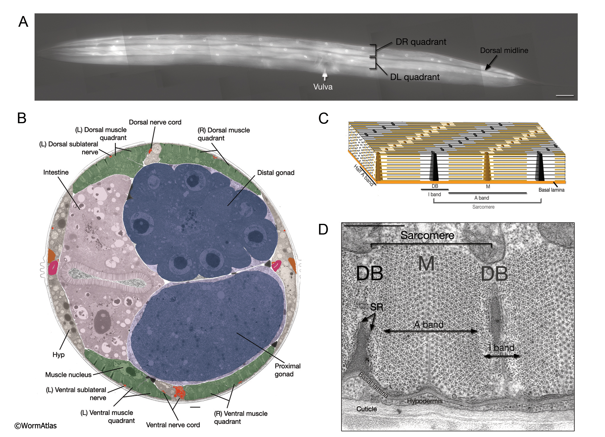
AMusFIG 1: C. elegans body wall muscles.
A. The organization of somatic muscles in the adult C. elegans hermaphrodite, dorsal oblique view, visualized using epifluorescence in a transgenic animal expressing the unc27::GFP reporter gene. The body wall muscles are organized in four quadrants (only the dorsal quadrants are visible here) with two rows of cells in each. The quadrants are placed subdorsally and subventrally. The two dorsal quadrants flank the dorsal hypodermal ridge and the dorsal cord, and the ventral quadrants flank the ventral hypodermal ridge and the ventral nerve cord. Anteriorly, the spindle-shaped cells in each quadrant are arranged almost in pairs, whereas more posteriorly the cells are organized in an alternating fashion. (Strain source: L. Jia and S.W. Emmons.) Bar, 50 µm.
B. Low-power TEM showing the relationship of the muscle quadrants to the hypodermis and internal organs, transverse section. Note the somewhat flattened profile of a normal muscle nucleus. Bar, 1 µm. (Image source: [Hall] N501-N354.)
C. Three-dimensional rendering of myofilament lattice as well as structure of the sarcomere. (Yellow lines) Thick filaments; (black lines) thin filaments; (black dots) dense body (DB); (brown dots) M lines.
D. TEM cross section of the contractile apparatus. The filaments of the lattice are oriented longitudinally in rows that are perpendicular to the surface. Dense bodies (DBs) anchor the thin (actin) filaments, whereas M line homologs anchor the thick (myosin) filaments. The membranous sacs of sarcoplasmic reticulum (SR) align around the dense body and are also present under the thick and thin filament bands along the muscle membrane. A single unit of myofilament lattice between two DBs is called a sarcomere and contains one A band in the middle and two juxtaposing half I bands. In C. elegans, each adult body wall sarcomere is about 1 µm wide. Bar, 0.5 µm. (Image source: [Hall] N501C.)
Click on picture for full resolution image.
|