|
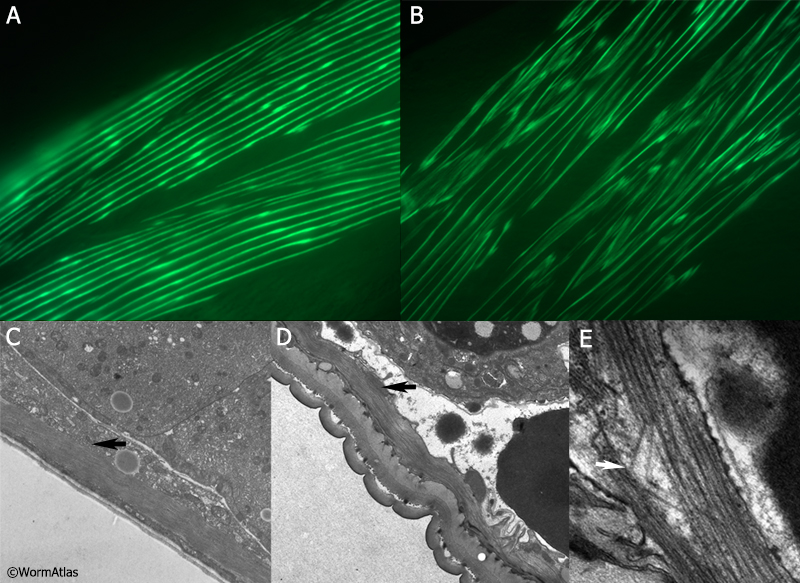
AMusFIG 2: Body wall sarcomeres in aging animals.
A&B. Muscle cell aging visualized using a MYO-3::GFP translational fusion highlighting the myosin heavy chain A in body wall sarcomeres from a young day 1 adult (A) and an aged day 15 adult, with evidence of sarcomere disorganization (B). (Image source: Herndon et al., 2002.)
C-E. Electron micrographs showing longitudinal organization of sarcomeres.
C. Muscle fibers in a young adult are evenly organized and are closely opposed to the contents of the muscle belly (black arrow). (Image source: N533 [Hall] 4235.)
D. Muscle fibers in older adults (15 days) are less cohesive and the muscle belly is grossly shrunken with few organelles seen (black arrow), so that the muscle cell plasma membrane (where it opposes the pseudocoelom) often seems collapsed against the sarcomere. (Image source: N815 [Hall] G713, Class B.)
E. In rare cases myosin filaments are frayed and bent (white arrow). (Image source: N803 [Hall] E769, Class C.)
Click on picture for full resolution image.
|