| PART/CELL NAME |
ABBREVIATION
SYNONYMS (S)
ANTONYMS (A) |
LINEAGE |
DESCRIPTION |
H1
H2 |
|
|
Head lateral ectoblasts which give rise to hypodermal cells in the head. |
| H band |
|
|
A well known feature of striated muscle in higher animals; a central, light staining region running in the middle of the A band. It is replaced by the “H zone” in C. elegans obliquely striated muscles.
See H zone
|
| “h”
neuron |
ASH cell (S) |
|
See ASH cell |
| H zone |
|
|
A thin dark line seen by polarized light running down the center of the A band in bodywall muscle sarcomeres. The A-band is very bright and contains interdigitating thick and thin filaments; the darker H zone contains only thick filaments (Miller et al., 1983; Waterston, 1988). |
| Habituation |
|
|
The reduction or absence of response after continued stimulation. This behavioral plasticity is commonly observed in C. elegans after repeated tapping of the head or tail with a hair or eyelash (See Rankin, 2005; Goodman, 2006). Similar reductions in response may also occur after prolonged exposure to chemical stimuli or other forms of stimulation (Rose and Rankin, 2001).
See Neural plasticity
|
| Half-desmosome |
Hemidesmosome (S)
Hemi-adherens junction (S) |
|
A term which is now archaic as molecular evidence suggests that the structure should be called a hemi-adherens junction.
See Hemi-adherens junction
|
| Harsh touch |
Hard touch (S)
Gentle touch (A) |
|
An aversive mechanical stimulus (e.g. poking the body with a metal wire), which is different than response to “gentle touch” (for instance with an eyelash to the bodywall). Harsh touch is transduced by the PVD sensilla in the back half of the body (Driscoll and Kaplan, 1997). Study of the genetics of sensory mechanotransduction have helped to elucidate the cellular and molecular components required to respond to harsh (and gentle) touch (Ernstrom and Chalfie, 2002).
See Touch response
|
| Hatchees |
|
|
Recently hatched L1 larvae. |
| Hatching |
|
|
A multi-step process, first described for C. elegans by Singh and Sulston (1978), by which the developing embryo makes an opening in the egg shell and exits this covering. Secretions from pharyngeal glands may promote he gradual weakening of the eggshell, which becomes visibly softer and more pliable in the hour just before eclosion. Once weakened, the motions of the three-fold embryo cause distortions of the whole shell and eventually the eggshell ruptures, allowing the new L1 larva to emerge from the broken shell.
See Eclosion
|
| Hatch-off |
|
|
A method for producing synchronized L1 progeny from an egg plate, either by picking individual maturing oocytes or by washing off hatching L1s as they get free of the eggshell. Eggs and eggshells tend to stick to an agar plate more avidly than a crawling L1 larva.
See Egg plate
See Lay-off
|
| Haustrulum |
|
|
A wide chamber within the pharyngeal lumen of the second bulb, immediately anterior and posterior to the grinder in some nematodes. It is not clear if this feature can be specifically identified in C. elegans (Nicholas, 1975). |
| Head |
|
|
A commonly used term in C. elegans literature that refers to the anterior end of the animal. This typically includes the anterior sensilla, pharynx and all muscles that are innervated by the nerve ring. Behind the head lies the body (or mid-body), including the intestine and reproductive tract. |
| Head lobe |
|
|
See Membranous organelle |
| Head mesodermal cell |
hmc |
|
A large single H-shaped muscle cell in the head that straddles the isthmus or second bulb of the pharynx and is anchored to the bodywall at the outside edge of each muscle quadrant and linked to these bodywall muscles by gap junctions (Sulston and Horvitz, 1977). Its exact function is unclear. |
| Head withdrawal |
|
|
A component of the foraging response, in which the animal quickly pulls its head away from a stimulus to the nose. The motion is mediated by the RMD motorneurons (Hart et al, 1995), and occurs faster than the typical rhythm at which the head swings back and forth in normal foraging.
See Foraging
|
| Healthspan |
|
|
The period of during adult life that is relatively free from age -related disease and physical impairment, which precedes senescent decline.
See Lifespan
|
| Heat avoidance |
|
|
See Thermal avoidance |
| Helminth |
Parasitic worm (S) |
|
Any species of parasitic worm, including flatworms, tapeworms, threadworms, and nematodes (roundworms). Usually the eggs of these worms lodge in the intestine of the host animal where they hatch, grown and multiply. |
| Hemi-adherens junction |
Hemidesmosome (S)
Dense body (S)
Half desmosome (S) |
|
A form of cellular attachment to extracellular matrices, homologous to the adherens junction, in which an asymmetric junction forms between a plasma membrane and a neighboring extracellular matrix (either basal lamina or cuticle). When attachments are to cuticle, there is often a corresponding dense specialization within the inner cuticle layer linked to the density on the plasma membrane. These junctions are common where hypodermis faces cuticle, and on the basal surface of some other epithelia, such as gonad sheath. The muscle dense body is probably a highly specialized form of hemi-adherens junction.
See Adherens junction
See Dense body
See Fibrous organelle
|
| Hemidesmosome |
Hemi-adherens junction (S)
Fibrous organelle (S)
Dense body (S)
Half desmosome (S) |
|
A term which is now archaic as molecular evidence suggests that the structure should be called a hemi-adherens junction. |
| Hemizonid |
|
|
A region of the ventral cuticle lying just posterior to the nerve ring that takes on a clear, lens-like appearance when viewed by light microscopy, apparently due to the close approach of the amphid commissure (McLaren, 1976).
See Amphid commissure
See Caudalids
See Cephalid
|
Hemizonion
|
|
|
A clear lens-like region of cuticle lying just posterior to the similar-looking hemizonid.
See Hemizonid
|
| Hermaphrodite/ Hermaphroditism |
|
|
The more common sexual form of the nematode, Caenorhabditis elegans, in which the gonad generates both male and female gametes. The hermaphrodite is self-fertile, since its gametes can be brought together internally to create healthy progeny. These animals can also participate in sexual unions with male animals to create cross-progeny by internal fertilization of the hermaphrodite’s female gamete, or oocyte, with the male’s sperm.
See Automictic
See Gonochoristic |
| Herniation |
|
|
A discrete bulging or local rupture in the basal lamina which allows a cell process to cross to the “wrong” side of a tissue border.
The same term might be used to describe a discrete bulging in any other thin membrane or tissue.
See Basal lamina
See Fenestration
|
| Hesitation |
|
|
A distinctive behavior noted when mutant animals must attend and compare two competing signals, decoded by distinct receptors in different neurons. Mutant hen-1 animals react normally to either stimulus alone, but stop moving briefly (hesitate) when presented with two competing signals, unable to overcome the aversive stimulus (Ishihara et al., 2002). |
| Heterotopy |
|
|
Variation in the relative position of an organ system from species to species. This term has been used with respect to the vulva position, which varies among nematode species in its position along the body axis (Fitch and Thomas, 1997; Sommer and Sternberg, 1994), but could also be used to compare the relative positions of sensilla or other body parts between species. |
| High frequency male producer |
|
|
A strain in which the spontaneous production of male progeny is increased above normal. In wild type C. elegans the normal production of males (due to spontaneous nondisjunction of the X chromosomes) is about 1 per 200 offspring; but him mutants may produce from 2-40% male progeny depending on the gene and allele. |
| High incidence of males |
him |
|
A strain with a mutant phenotype in which the X chromosome is spontaneously lost at a relatively high rate during meiosis in the germline. This loss changes the normal XX karyotype of hermaphroditic progeny to XO, resulting in more males and fewer hermaphrodites (Hodgkin et al., 1979). Generally, these males are fertile and appear normal in appearance and behavior when compared to spontaneous males.
See Andric index
See Spontaneous male
|
| Hindgut |
|
|
The more posterior portion of the intestine, just anterior to the sphincter valve, which is subject to squeezing by the somatal-intestinal muscles. |
| Histology |
|
|
The microscopic study of tissues to gain an understanding of the cellular and subcellular features of animal or plant life. |
| Holidic medium |
|
|
A chemically defined culture medium, in which every component is well-defined. A defined medium containing one less well-characterized component, such as a cell extract, is termed a meridic medium.
See Axenic medium
|
| Holocentric chromosomes |
|
|
A normal feature of C. elegans chromosomes (as opposed to some other animal species which have “monocentric” chromosomes). During chromosome duplication at metaphase, monocentric chromosome pairs appear to be attached at only one position along their length, whereas “holocentric” chromosomes appear to line up in parallel and be closely attached at multiple positions (Albertson and Thomson, 1982; White, 1973; Albertson et al., 1997). This property may be important in allowing the nematode to accurately segregate small chromosomal fragments (free duplications) to both daughter cells with minimal error, but also seems to facilitate a much higher level of chromosomal rearrangements (Dernburg, 2001; Coghlan, 2006).
See Kinetochore
See Monocentric chromosomes
|
| Holomyarian |
|
|
A condition in which a nematode muscle quadrant contains only 1 or 2 cells across, as opposed to “meromyarian” muscles which contain 2-5 cells across (as in C. elegans), or “polymyarian” muscles which contain greater than 5 cells across within a quadrant (Schneider, 1860; Chitwood and Chitwood, 1950; Bird and Bird, 1991).
See Meromyarian
See Platymyarian
|
| Homeostasis/ Homeostatic |
|
|
The ability of an animal to regulate its internal environment to create stability despite external forces that might otherwise be either toxic or disruptive to normal growth and development. |
| Hook |
|
|
A small lump on the gubernaculum of the male tail. This structure lies on the ventral surface just anterior and central to the base of the spicule openings and contains a sensory structure called the hook sensillum.
Alternately, can refer to a curved extension protruding from some doublet microtubules in sensory axonemes which may act to connect parallel microtubules to their neighbors or to the plasma membrane of the cilium. |
| Hook sensillum |
Anterior sensillum (S) |
|
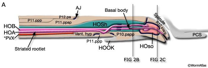
Two specialized sensory neurons (HOA and HOB) enclosed by a sheath cell (HOsh) and socket cell (HOso) on the ventral surface of the gubernaculum in the male tail. A narrow channel to the exterior suggests that the neurons could have chemosensory function. The hook points anteriorward, whereas the paired post-cloacal sensilla point in posteriolateral directions just behind the hook and cloacal opening. |
| Host |
|
|
The animal or plant species in which a parasitic species (of nematode) receives its nutrition or shelter, and in which it must infect prior to reproducing itself. Some species may require several different hosts in order to advance through a complete life cycle.
See Life history
See Parasite |
| Host finding |
|
|
A prominent behavior in parasitic nematodes which is necessary for completion of their life cycle. Some Rhabditid nematodes have a commensal lifestyle in the wild to live under the shells of isopods or snails, including C. remanei and C. vulgaris (Baird et al., 1994; Baird, 1999). In such cases the nematode may utilize chemosensory circuits to identify and move towards one or more host species. |
| Host/ Pathogen interaction |
|
|
This term can refer to either the success rate of infectivity (pathogen entering the host), or the success rate of killing (pathogen killing the host).
See Hypervirulent
|
| HSN |
HSNL
HSNR
Hermaphrodite specific neuron (S) |
ABplapppappa
ABprapppappa |
An isolated cell pair lying in the midbody of the adult that act to operate the vulval musculature during egglaying. |
| Hyaline sphere |
Yolk granule |
|
|
| Hydrostatic skeleton |
Hydroskeleton (S)
|
|
High internal pressure maintained within the pseudocoelom, pushing outward on the cuticle, can give the nematode a significant structural integrity not unlike the solid skeleton in higher animals. In fact, C. elegans is known to be able to leave the substrate and actively wave its body in the air (nictation), holding fast just by its tail.
|
| Hypercontraction phenotype |
|
|
A class of muscle mutant (such as unc-90) in which the body displays a gross shortening and stiffness due to general contraction of all bodywall muscles at once. |
| Hyperoxia |
|
|
A high level of oxygen in the environment (13-21% O2), which the nematode finds stressful and aversive. The animal will use aerotaxis to move back to lower oxygen levels, and will become more social and show more “bordering” behavior on culture plates (Gray et al., 2004). |
| Hypervirulent |
|
|
Refers to bacterial strains which are exceedingly toxic when ingested by C. elegans (Mylonakis et al., 2002).
See Host/ pathogen interaction
See Killing assay
|
| Hypodermal cell |
|
|
Very few hypodermal cells maintain separate identities after morphogenesis in the embryo as most fuse into larger hypodermal syncytia almost immediately (Podbilewicz and White, 1994). Exceptions to this rule include hyp 8, hyp 9 and hyp 11 in the tail (Nguyen et al., 1999). The same rule holds true for later larval lineages, where additional hyp cells are born, begin to differentiate and then fuse into local syncytia.
See Hypodermal syncytium
|
| Hypodermal ridge |
Hypodermal cord (S)
Hypodermal line (S) |
|
The set of longitudinal swellings of hypodermal tissue that develop during elongation phase of embryogenesis and persist throughout the life of the animal. Hypodermal tissue becomes squeezed aside as the four bodywall muscles quadrants settle against the cuticle resulting in a series of four distinct rows of hypodermal tissue in two wide lateral ridges and thinner ventral and dorsal ridges of hypodermis along the full length of the larva and adult. A fifth anal ridge of hypodermis extends posteriorly into the tail tip. Within each ridge are the nuclei of the hypodermal syncytium, while very thin hypodermal extensions wrap underneath each row of muscle cells to connect all the ridges together.
See Cord/ Chord
See Lateral line
|
| Hypodermal segmentation |
|
|
A series of hypodermal syncytia adopt restricted circumferential territories along the length of the body, including 5 sets of syncytia occupying narrow territories in the lips, hyp 6 covering a portion of the head and hyp 7 covering most of the length of the body. The posterior tail is not organized in the same circular territories, but one can still see a rough segmentation, numbering anteriorly to posteriorly hyp 8 + hyp 12, hyp 9 + hyp 11 and hyp10. |
| Hypodermal syncytium |
|
|
The characteristic fate of hypodermal cells is to quickly fuse to create larger syncytia, most of which encircle a given segmental region along the length of the body. Their cell bodies and most cytoplasm are clustered in hypodermal cords along the lateral, ventral and dorsal sides, connected by very thin cell extensions running beneath the muscle quadrants, always closely apposed to the body cuticle. Virtually all syncytia lie within the bodywall, comprising the most peripheral tissue covering the whole body, eg. the ectoderm. |
| Hypodermis |
Ectodermis (S)
Epidermis (S) |
|
The accepted term for the primary epidermis in nematodes (Chitwood and Chitwood, 1950; Bird and Bird, 1991), consisting of a single layer of cells directly underlying (and secreting) the cuticle. Most of the hypodermal cells become syncytial during embryogenesis, so that the hatched larva is covered by just a small number of hypodermal syncytia, some having over 100 nuclei. Several forms of interfacial (transitional) epidermal cells are also considered part of the hypodermis, including the seam cells, the socket cells and those cells specialized to secrete the cuticular linings of the buccal cavity, vulva and rectum. |
| Hypodermoblast |
|
|
An undifferentiated ectodermal cell that is fated to yield a mature hypodermal cell, or to divide to produce accessory neurons, glial cells and one hypodermal cell. |
| Hypoxia/ Hypoxic |
|
|
A very low level of oxygen in the culture medium (0.5%-4% O2), under which C. elegans shows reduced activity and sometimes death (Epstein et al., 2001; Jiang et al., 2001). Survival from long term hypoxia seems to involve different molecular mechanisms than those required for survival from complete anoxia (Padilla et al., 2002).
See Anoxia
See Cryptobiosis
See Hyperoxia
See Normoxia
|

