|
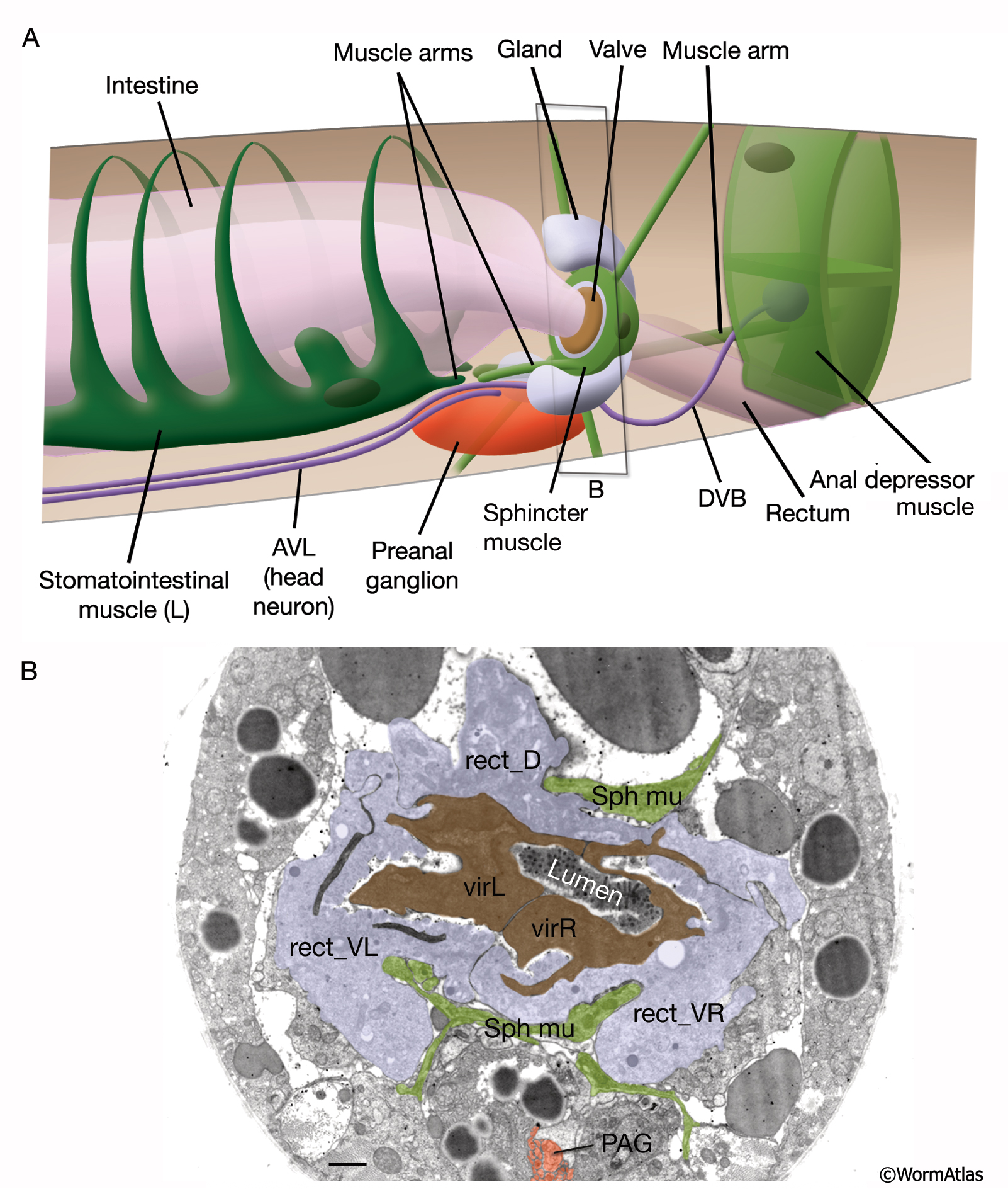
RectFIG 1: The hindgut.
A. Graphic rendition of the structures of the posterior alimentary canal. (Dark ovals) Muscle nuclei.
B. The lumen of the posterior intestine ends at the rectal valve, composed of two cells (virL and virR). The valve is surrounded by a trilobed rectal gland composed of three cells (rect_D, rect_VL, rect_VR). The gland cells connect to the lumen just posterior to the valve (not shown). The sphincter muscle (Sph mu) surrounds and pierces into the gland. TEM, transverse section. (Image source: [Hall] B140A-T581.) Bar, 1 μm.
Click on picture for full resolution image.
|