|
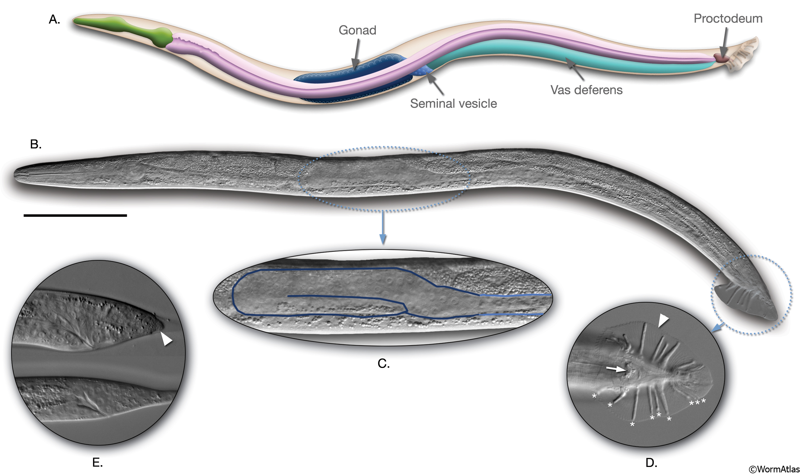
IntroFIG 5: C. elegans male.
A. Schematic drawing of anatomical structures, left lateral side.
B. DIC image of an adult male, left lateral side. Scale bar 0.1mm.
C. The unilobed distal gonad of the animal in B is shown as enlarged.
D. The adult male tail, ventral view. (Arrow) Cloaca; (arrowhead) fan. Rays 1-9 are labeled with asterisks on the right side.
E. L3 tail, bottom, starting to bulge. The tail hypodermis has retracted in the L4 tail (arrowhead), top (compare with IntroFIG8).
Click on picture for high resolution image. |