|
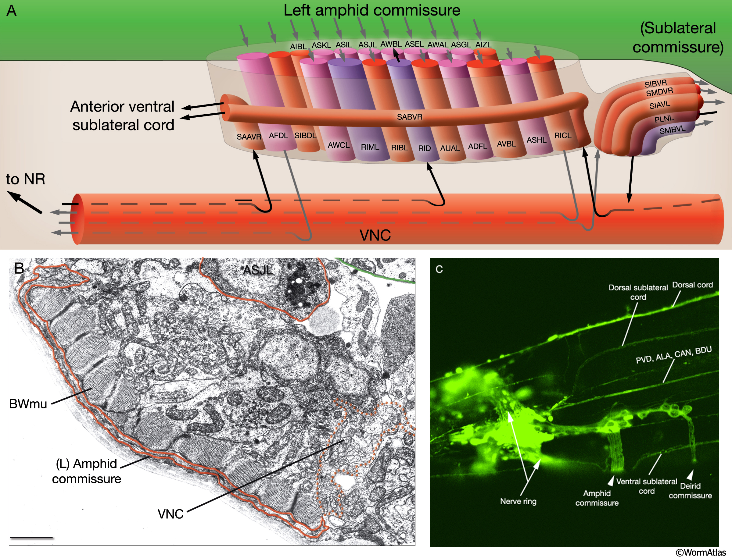
NeuroFIG 14: The composition of the left-side amphid commissure.
A. Schematic of the commissure, left lateral view. The axons of the amphid neurons enter the VNC through the ipsilateral amphid commissure en route to the NR. (The trajectory of the RIBL axon within the commissure is shown in more detail in NeuroFIG 15.) The ventral process of RID, a single neuron situated in the dorsal ganglion, runs down stochastically on one side of the NR and enters the VNC. It then exits the VNC via the same-side amphid commissure and continues as a commissure to reach the DC. The processes of SABV and SAAV join the anterior subventral cords through the amphid commissures. The posterior part of the amphid commissure, which is also referred to as the sublateral commissure, consists of the processes that are entering or exiting the ventral sublateral cords. (Based on White et al., 1986.)
B. TEM of the left amphid commissure, transverse view. Also shown is one of the neurons that sends an axon to the commissure, ASJL. Processes travel between the muscle and hypodermis, underneath the hypodermal basal lamina. The VNC is split into two approximately equal thickness bundles at this level, split by the excretory pore and ventral hypodermal ridge. The processes from the left-sided commissure join the left side of the VNC (dotted red line). Bar, 1 μm. (Image source: [MRC] N2T-101-15.)
C. Confocal image from transgenic animals expressing unc-104::GFP reporter gene showing longitudinal and circumferential fiber tracts and commissures in the head, lateral view (See also NeuroFIG 19A). (Image source: O. I. Wagner. Image taken by Wu Gong-Her on an Olympus FV1000 at NSRRC, Hsinchu, Taiwan; strain generated by D.R. Klopfenstein.)
Click on picture for full resolution image.
|