|
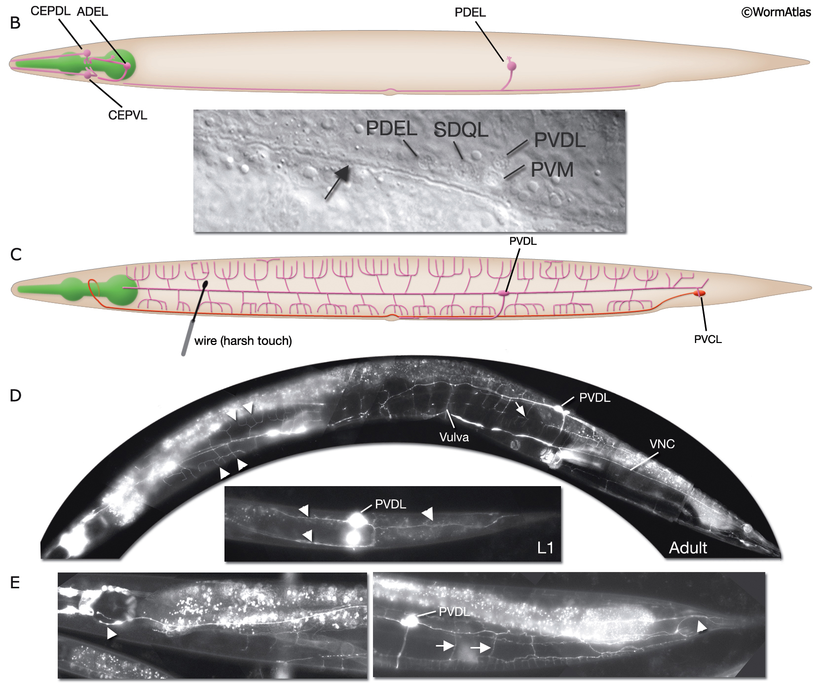
NeuroFIG 10B-E: Sensory neurons that sense nose touch, osmotic pressure, texture, and harsh touch.
In all panels, only the cells on the left side are shown.
B. Dopaminergic neurons that detect stimulus texture. Each neuron has a ciliated ending either in the head or midbody. (Top) Schematic rendition, lateral view. (Bottom) DIC image of the posterior lateral side of the body. The nuclei of the neurons in the left lateral ganglion are seen close to the excretory canal (arrow). Magnification, 600x.
C. Graphic rendition of neurons required for perception of harsh touch to the body (PVD) and tail (PVC), lateral view.
D&E. PVD neurons. D. (Top panel) Adult, ventral-lateral view. PVD neurons have multiple short branches (arrowheads) that arise at the level of the muscle quadrants (arrow). (Bottom panel) L1 animal, left lateral view. Although the three main processes are already formed (arrowheads), few branches are seen. E. (Left panel) Adult, lateral view. PVD processes terminate around the region of the posterior bulb of the pharynx (arrowhead). (Right panel) Adult, lateral view. Multiple branches arise from the posteriorly directed PVD process (arrows), which continue to the tail region (arrowhead). All are epifluorescent images of transgenic animals expressing the ser2prom3::GFP reporter in PVD neurons on a roller background. Magnification, 400x. (Strain source: O. Hobert.)
See also NeuroFIG 10A
Click on picture for full resolution image.
|