|
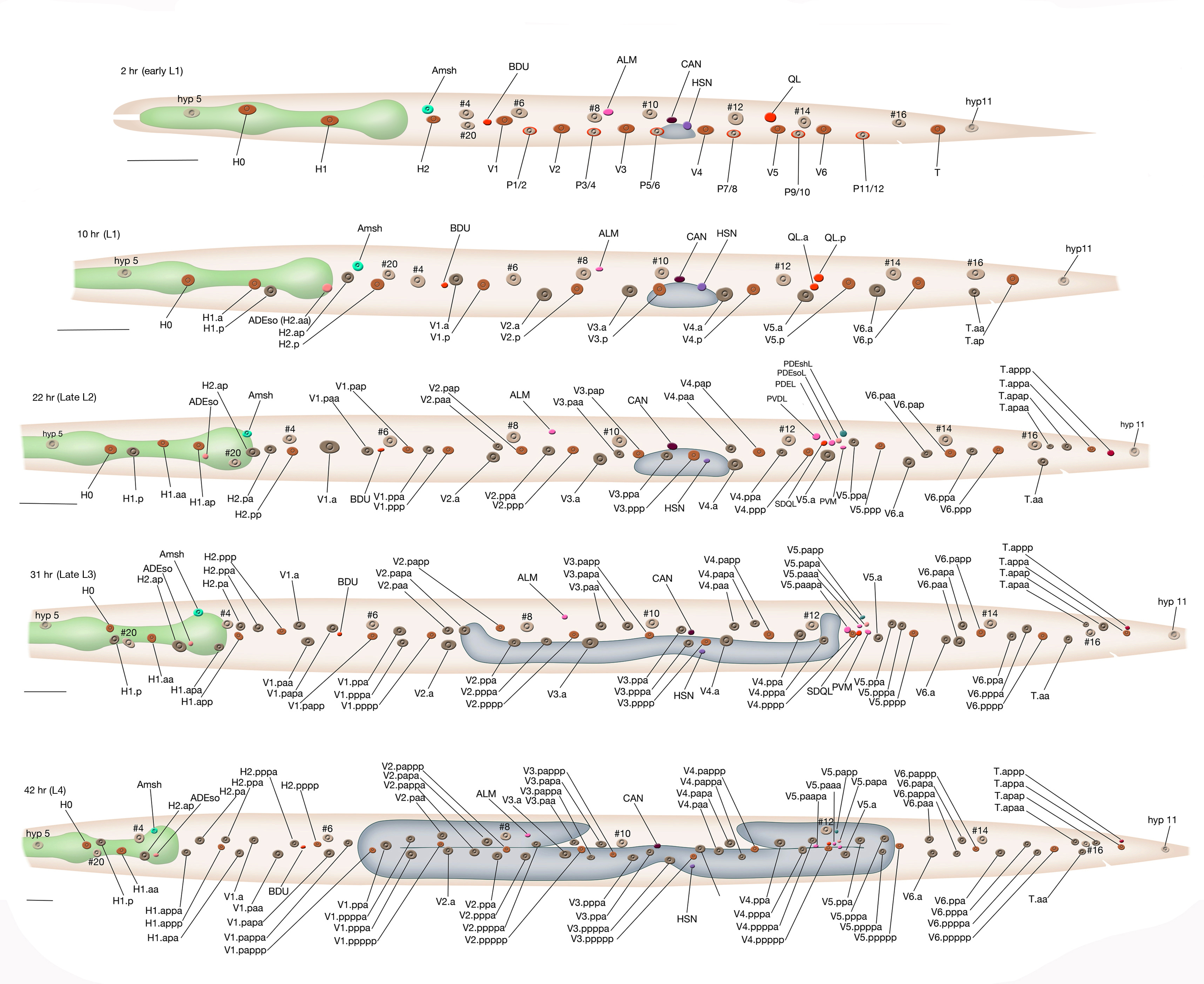
HypFIG 8G: Graphic depiction of the postembryonic development of the left lateral hypodermis.
Bars on each panel correspond to 20 μm. The number signs next to some of the hyp nuclei correspond to those in HypFIG 2 panel D. These are the embryonically-derived hyp nuclei and are colored as light beige, while postembryonically-born nuclei are colored darker. The colors of the remaining nuclei correspond to the WormAtlas color code. The names of the postdeirid ganglion nuclei are shown only in the third (22 hr) panel. Based on Fig 9 in Sulston and Horvitz, 1977.
Click on picture for full resolution image.
|