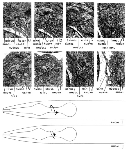
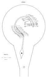
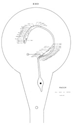
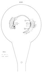
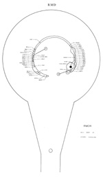
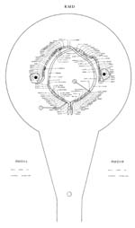
RMD is a set of six major motoneurons, which innervate muscles in the head via NMJs in
the nerve ring. The cell bodies of RMDV and RMD are situated in the lateral ganglia adjacent
to the ring neuropile; those of RMDD are in the ventral ganglion (figure 16). Processes enter
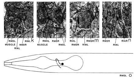
the nerve ring laterally in the case of RMDV (j) and RMDL/RMDR (o), and via the ventral cord
in the case of RMDD (i), and run round the ring for three quadrants. The proximal regions
of these processes run closely associated together and are also closely associated with, and often
surround, the processes of RIA (d). The processes of this group run in the middle of the anterior
regions of the neuropile of the ring. The distal regions of each of the RMD processes move
towards the inside surface of the ring, where they have their NMJs. RMDD wraps round the
process of CEPD in a characteristic manner (g). The main regions of synaptic input are
diametrically opposite the regions of NMJs (the NMJs present on the left-hand side of RMDL are atypical and have not been seen on the other reconstructed series).
The NMJs of RMDD/RMDV are usually in a complex that consists of NMJs from RMD, IL1 and URA and dendrites from RMD and RIP (a, b). Sometimes the complex is seen without
the RIP and URA processes
(c). RMDL/RMDR behave somewhat differently and could, possibly, be considered as a separate
class. Most of the NMJs of RMDL/RMDR are part of a complex, present on each of the four muscle
arm spurs (figure 14), in which an RMDL/RMDR dendrite is
sandwiched between an RMDL/RMDR NMJ and an RIM NMJ (k). There are also a few RMDL/RMDR NMJs laterally that are not part
of any complex (I). The processes of RMDL/RMDR flatten out and form caps over the anterior
surfaces of the NMJ regions of RIM, which are situated directly anterior to the muscle arm
spurs (m, RIM-*b, RIM-*c). The main synaptic outputs of RMDL/RMDR are onto contralateral RMDL/RMDRs (as corecipients at NMJs) and RIA (n); the main synaptic inputs are: RIA (*e), RIM (m), RMG (*a), ILl, ADE (*c), RMF (*e),
contralateral RMDL/RMDRs, RIS (*d), RMH (*a)
and OLL (d). There are also gap junctions to RMDD/RMDV, but not to each other. The only
synaptic output of RMDD/RMDV, apart from NMJs, is to contralateral RMDD/RMDVs at the NMJ
complexes; the main synaptic inputs are from RIA (*f), URY (f), contralateral RMDD/RMDVs
(a, b), IL1 (a, b), OLL (*d), CEP (*h), RIV (*d) and OLQ (*b). There are gap junctions
to SAA, RMDD/RMDV, SMD, RMDL/RMDR, AVE and OLQ. OLQ has an interesting
complementary synaptic relation to RMDD/RMDV; OLQD synapses onto RMDD and has gap junctions
to RMDV, whereas OLQV synapses onto RMDV and has gap junctions with RMDD.
Occasionally the processes of RMDD/RMDV appear to penetrate the basal lamina separating
nervous tissue from muscle arms, and have gap junctions directly onto muscle arms (h).
Magnifications: (a, h, l) x 25500, (b, c, e-g, k, m, n) x 12750, (d) x 17000.

Click pictures for higher resolution images
