Click pictures for higher resolution images Click pictures for higher resolution images
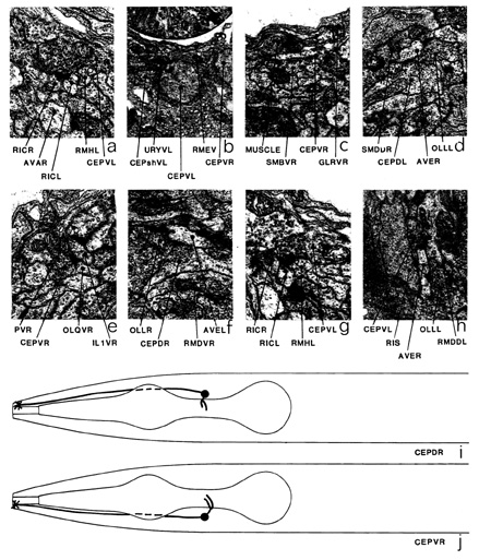
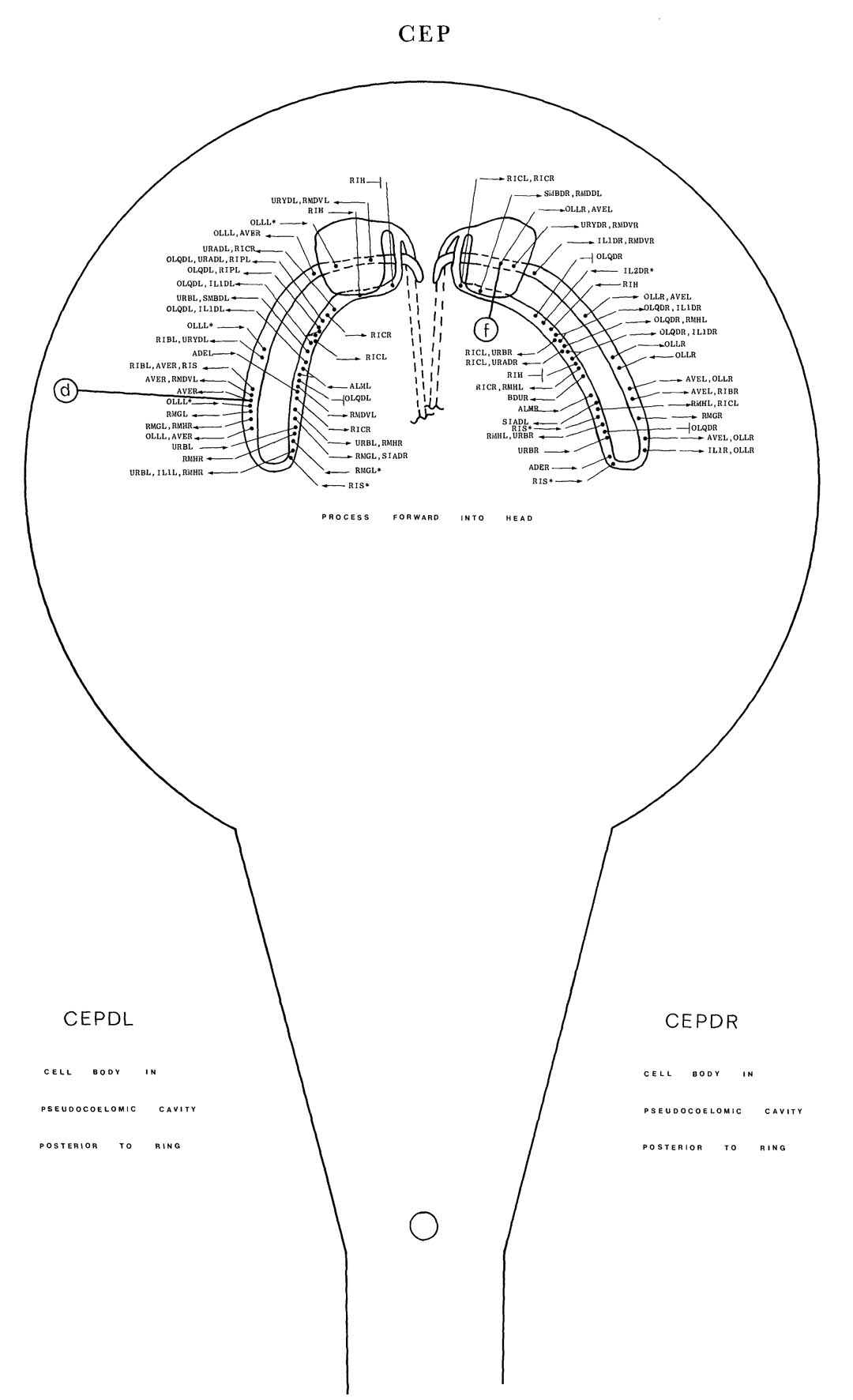
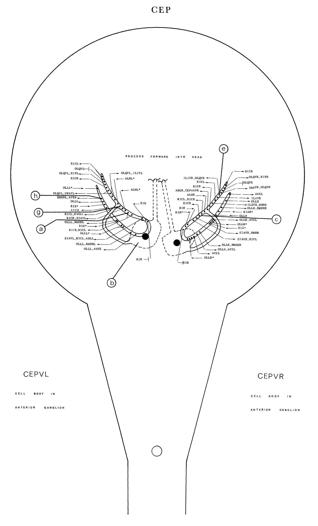
CEP is a set of four neurons with ciliated endings in the cephalic sensilla (figure 1). The
dorsal pair of cell bodies is situated in the pseudocoelomic cavity posterior to the nerve ring
(along with those of URX). The ventral cell bodies are situated anterior to the ring and closely
apposed to the ring neuropile (b). Anteriorly directed processes run in four of the six labial
process bundles to the receptor endings in the head. Posteriorly directed processes emanate
from the ventral CEP pair and loop round the posterior face of the ring
neuropile; they then enter it on the inside surface adjacent to the muscle arms (j, c). The processes branch at this
point and run both ways round the nerve ring on the inside surface near the posterior face of
the ring neuropile. The dorsal branch ends; the ventral branch loops round and runs dorsally
in the middle of the anterior regions of the ring neuropile. The dorsal pair of CEP neurons
send out anteriorly directed processes, which enter the ring near the dorsal mid-line (i) and
then run ventrally on the inside of the ring neuropile adjacent to the muscle arms. These then
loop back and run in the middle of the anterior regions of neuropile, eventually moving back
to the inner surface, where they end. The main synaptic output is to RIC (a, g), AVE (d, f,
h), OLL (f), OLQ (e), ILl (e), RMH (g), RMD (h), RMG, URA and URB. CEP synapses
have been shown to contain the neurotransmitter dopamine (Sulston et al. 1975). There is some
synaptic input from OLL, ALM (*c), RIH, RIS, and also from URB (*b) and ADE to the
dorsal pair only. There are gap junctions to OLQ (*e) and RIH.
Magnifications: (a, e, g) x 25500, (b) x 6375, (c, d, f, h) x 12750.

|
|