|
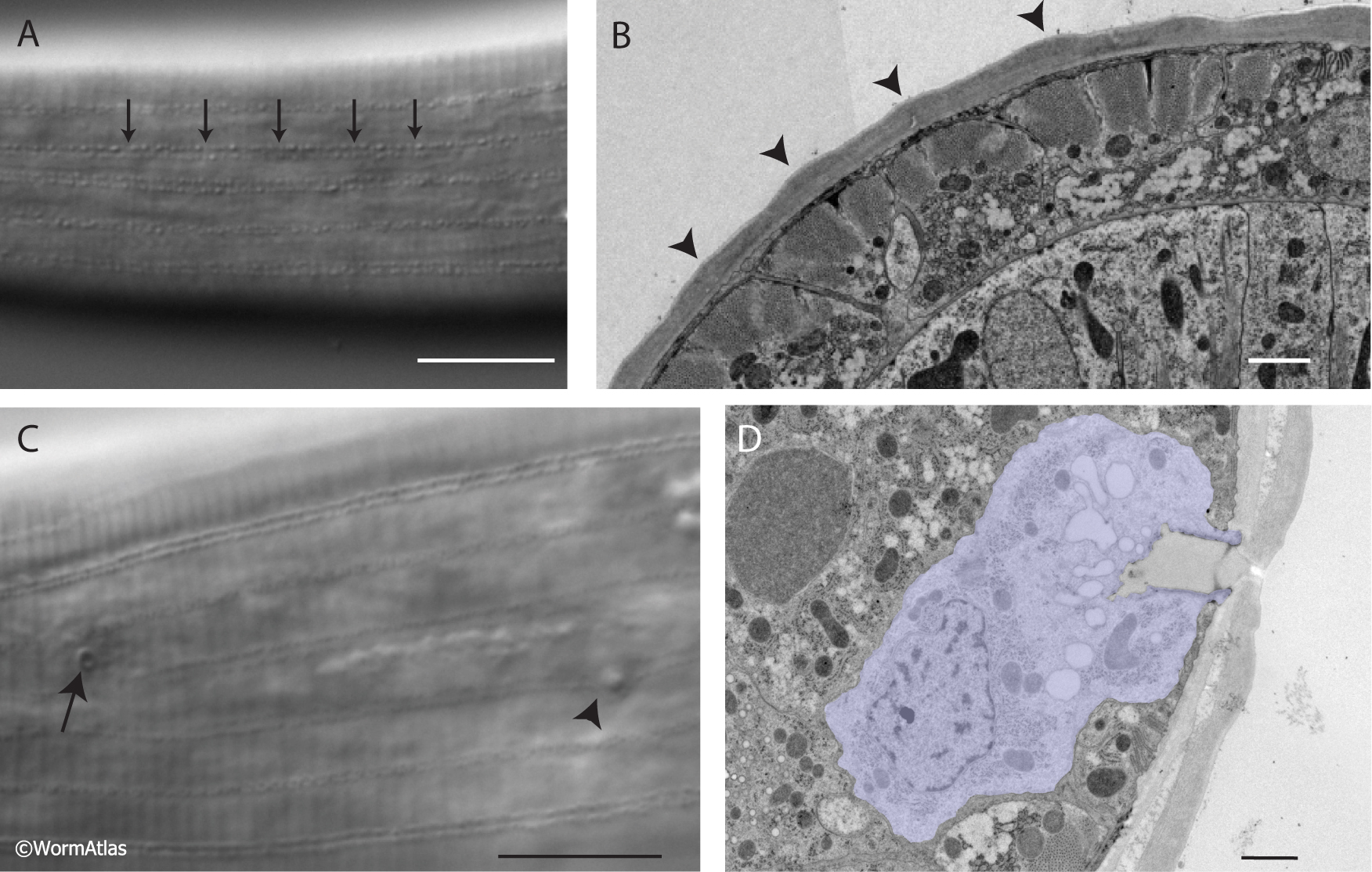
PIntroFIG 4: The cuticle of P. pacificus.
A. The cuticle of P. pacificus contains longitudinal stippled lines around the circumference of the animal. Scale bar, 10 um.
B. In an EM cross section, this stippling can be seen as waves around the animal (arrowheads). Scale bar, 1 µm. (Image source: Ralf Sommer Lab, Bumbarger13-1301.)
C&D The cuticle of P. pacificus is interrupted by epithelial gland cell pores. In DIC (C), the small gland cell pores (arrow) are occasionally visible both dorsal and ventral of the midline (deirid, arrowhead). Scale bar, 10 µm. In EM cross sections (D), the epithelial gland cell (purple) is surrounded by the hypodermis. Scale bar, 1 µm. (Image Source: Ralf Sommer, Bumbarger14-1955.)
Click on picture for full resolution image.
|