PhaMOVIE 1A. Development of a 3-D pharynx. Ventral view. Pharynx is derived from AB and MS lineages (Santella et al, 2010). 0 min is first cleavage (which is approximately at 40 min after fertilization). Colors do not follow the WormAtlas color code. Click on the movie to open a larger version in a separate page (Movie source: A. Santella, J. Moore and Z. Bao.)
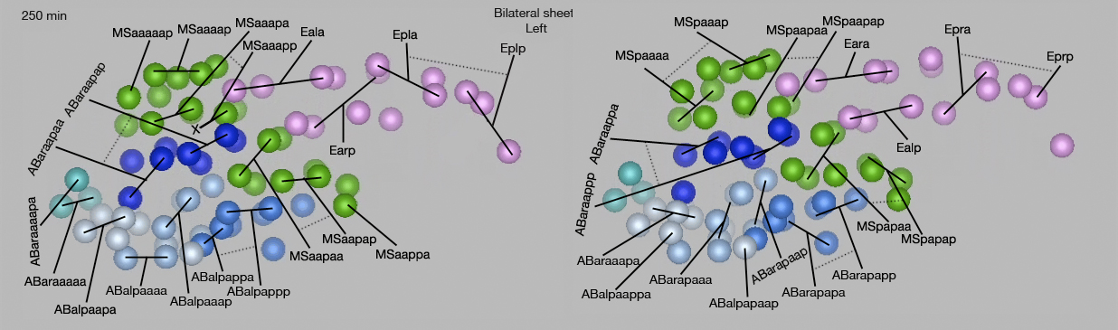
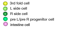
Lineage Key at 250 min for PhaMOVIE 1A&B Lineage relationships of cells that make up the bilateral sheet of the pharynx primordium at 250 min after first cell cleavage. Top row: ventral view, middle row: left side view, bottom row: dorsal view. Dotted lines indicate sister cells. X indicates programmed cell death. Click on images for higher resolution versions.
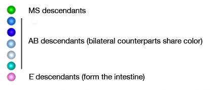
Lineage Key at 337 min for PhaMOVIE 1A&B Each cell is labelled with its lineage name. (Blue spheres) Cells from AB lineage; (green spheres) cells from MS lineage; (pink spheres) cells from E lineage that form the intestine. Colors do not follow the WormAtlas color code. Click on the movie to open in a separate page (Movie source: A. Santella, J. Moore and Z. Bao.)
PhaMOVIE 1B. Development of a 3-D pharynx. Left side view. Pharynx is derived from AB and MS lineages. 0 min is first cleavage (which is approximately at 40 min after fertilization). (Blue spheres) Cells from AB lineage; (green spheres) cells from MS lineage; (pink spheres) cells from E lineage that form the intestine. Colors do not follow the WormAtlas color code. Click on the movie to open a larger version in a separate page(Movie source: A. Santella, J. Moore and Z. Bao.)
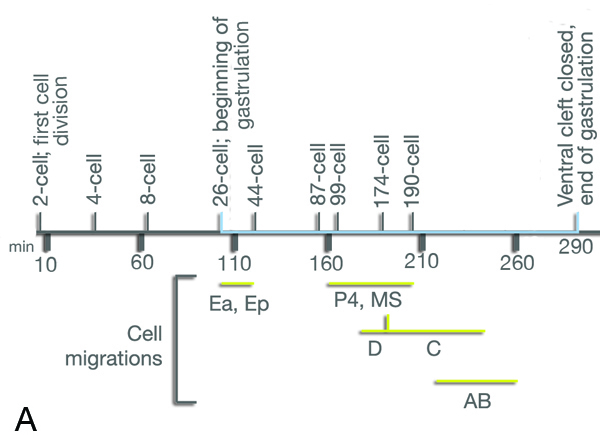
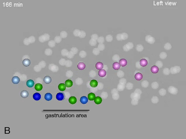
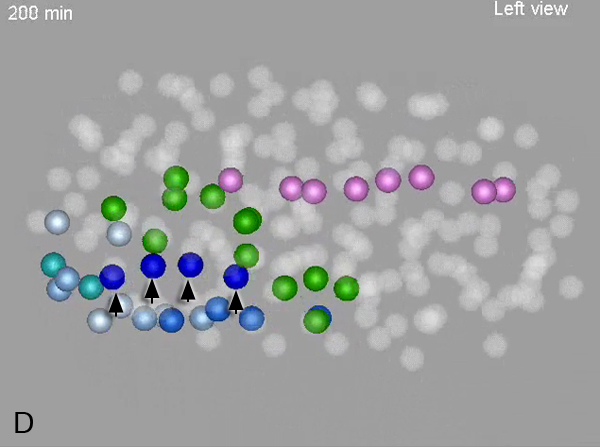
Gastrulation of the pharyngeal cells. A. Gastrulation time course. Time is indicated in minutes below the bar. 0 time point is first cell division which takes place about 40 min after fertilization at 20oC. Blue bar indicates gastrulation phase. By 120 min (just before the E2-4 division) movement of gut precursor Ea and Ep cells to inside the embryo is completed. B. Around 160 min MS cells start to move in via the gastrulation cleft (indicated with a black bar). C. Around 190 min MS cells complete their movement (indicated by arrows) to inside the embryo and the AB cells now cover the ventral surface. D. Around 200-210 min AB cells start to move inside the embryo (indicated by arrows) via the cleft.
By 260 min all pharyngeal cells reside inside the embryo (not shown). B-D: (Blue spheres) Cells from AB lineage; (green spheres) cells from MS lineage; (pink spheres) cells from E lineage.
2 Recruitment of cells between the layers of the bilateral sheet to create a spherical pharynx
PhaMOVIE 2. Formation of the 3rd layer. Ventral view. After a bilayer of pharynx primordium forms at about 250 min, cells from both AB and MS lineages wedge themselves between this bilayer to create a 3-D pharynx (for development of 3-fold symmetry, a third, equivalent cell joins a bilateral pair and pharynx rounds at around 335 min). 0 min is first cleavage (which is approximately at 40 min after fertilization). Colors do not follow the WormAtlas color code. Click on the movie to open a larger version in a separate page (Movie source: A. Santella and Z. Bao.)
Lineage and spatial relationships of 3rd layer cells at 250 min. Cells that contribute to the 3rd fold of the pharynx (yellow spheres) do not seem to show any lineage-based bias or regularity as they give rise to a mixed variety of cells, including pharyngeal neurons, muscle, epithelium, mc cells, pharyngeal-intestinal valve and arcade cells. Rather, they seem to rely on a spatial logic such that they are born at or near the midline along the A-P axis of the embryo (Santella et al, 2010). Terminal cells that are born from each 3rd-fold precursor are listed in parenthesis underneath the lineage name of the precursor. (Dark green spheres) Left sheet of the bilateral pharynx primordium; (light green spheres) right sheet of the bilateral pharynx primordium.
by Santella, A.*, Bao, Z., Moore, J.,Hall, D.H. and Altun, Z.F.* 2012.
*to whom correspondence should be addressed: santella@mskcc.org; zeynep.altun@einstein.yu.edu