|
Pharynx Atlas Home
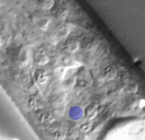
The pictures that comprise the Pharynx Atlas are a focal series through the terminal bulb of the
pharynx. The worm is lying on its right side, with anterior towards the upper left and posterior towards the lower right. Dorsal is the upper right and ventral is the lower left.
This is the fourth image in the series.
Earlier images start at the leftmost edge of the pharynx and go consecutively deeper, until the last goes through
the very rightmost edge. The focal planes are not equally spaced. They
were chosen so that all the nuclei in the terminal bulb would be
clearly visible in at least one image.
Click on individual pharyngeal cells to identify them.
Pharyngeal Gland Cell g2L
Description: The two g2 gland cells fill the ventral sector of the most posterior region of the terminal bulb. They extend processes anteriorly to open on the lumen of the terminal bulb. Their function is unknown. (See Pharynx section; Albertson and Thomson, 1975.)
Pharynx Focal Plane 4
|
Identification: The g2 nuclei have a very characteristic appearance: small, very nearly spherical, with a smooth nucleoplasm and a small nucleolus usually near the exact center. They are side by side on the ventral midline of the terminal bulb, just posterior to mc3V. |
|
