|
Pharynx Atlas Home
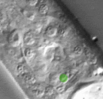
The pictures that comprise the Pharynx Atlas are a focal series through the terminal bulb of the
pharynx. The worm is lying on its right side, with anterior towards the upper left and posterior towards the lower right. Dorsal is the upper right and ventral is the lower left.
This is the third image in the series.
Earlier images start at the leftmost edge of the pharynx and go consecutively deeper, until the last goes through
the very rightmost edge. The focal planes are not equally spaced. They
were chosen so that all the nuclei in the terminal bulb would be
clearly visible in at least one image.
Click on individual pharyngeal cells to identify them.
Pharyngeal Terminal Bulb Muscle Cell pm8
Description: pm8 is a small toroid muscle at the very back of the terminal bulb. It is attached to the pharyngeointestinal valve, and may operate it. (See Pharynx section; Albertson and Thomson, 1975.)
Pharynx Focal Plane 3
|
Identification: The pm8 nucleus is very hard to find. It is a small fried egg near the axis of the pharynx and at the extreme posterior end. |
|
