|
Pharynx Atlas Home
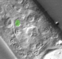
The pictures that comprise the Pharynx Atlas are a focal series through the terminal bulb of the
pharynx. The worm is lying on its right side, with anterior towards the upper left and posterior towards the lower right. Dorsal is the upper right and ventral is the lower left.
This is the third image in the series.
Earlier images start at the leftmost edge of the pharynx and go consecutively deeper, until the last goes through
the very rightmost edge. The focal planes are not equally spaced. They
were chosen so that all the nuclei in the terminal bulb would be
clearly visible in at least one image.
Click on individual pharyngeal cells to identify them.
Pharyngeal Terminal Bulb Muscle pm5VL
Description: The six pm5 nuclei belong to three muscle cells, each of which runs the length of the isthmus. The muscle cells are dorsal (pm5DL and pm5DR), left subventral (pm5L and pm5VL), and right subventral (pm5R and pm5VR), and are separated from each other by the mc2 marginal cells. (See Pharynx section; Albertson and Thomson, 1975.)
Pharynx Focal Plane 3
|
Identification: All the pm5 nuclei are large fried eggs. The two pm5V nuclei are found in the ventral anterior terminal bulb just anterior to I5. The two ventral nuclei are found in the ventral anterior terminal bulb just anterior to I5. They are very close together on either side of the ventral midline. The lateral and dorsal pm5 nuclei are at the same A/P level of the TB. Together with g1A, they form a triangular cluster in the subdorsal anterior terminal bulb, dorsal to the pm5V nuclei. The ventral and dorsal nuclei are anterior of g1A, which is anterior ventral of a neuron (M1 on the right, I6 on the left). The three nuclei are not usually visible in the same focal plane. |
|
|
