|
Pharynx Atlas Home
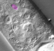
The pictures that comprise the Pharynx Atlas are a focal series through the terminal bulb of the
pharynx. The worm is lying on its right side, with anterior towards the upper left and posterior towards the lower right. Dorsal is the upper right and ventral is the lower left.
This is the third image in the series.
Earlier images start at the leftmost edge of the pharynx and go consecutively deeper, until the last goes through
the very rightmost edge. The focal planes are not equally spaced. They
were chosen so that all the nuclei in the terminal bulb would be
clearly visible in at least one image.
Click on individual pharyngeal cells to identify them.
Pharyngeal Marginal Cell mc2DL
Description: The three mc2 marginal cells run the length of the isthmus in ventral, left subdorsal, and right subdorsal sector. They are structural cells that anchor the apices of the triradiate lumen. Inside they have intermediate filaments that run from the lumen apex to the basement membrane that surrounds the pharynx. (See Pharynx section; Albertson and Thomson, 1975.)
Pharynx Focal Plane 3
|
Identification: The mc2 nuclei are the most anterior in the anterior terminal bulb, or perhaps even in the posterior isthmus. mc2V is on the ventral midline anterior to the g1A nuclei. mc2dr and mc2DL are anterior to the pm5 nuclei. They have one or two prominent nucleoli. Often the nuclei have a characteristic bilobed shape, probably because they are indented by the apices of the pharyngeal lumen. |
|
