|
Pharynx Atlas Home
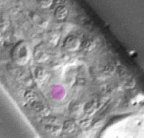
The pictures that comprise the Pharynx Atlas are a focal series through the terminal bulb of the
pharynx. The worm is lying on its right side, with anterior towards the upper left and posterior towards the lower right. Dorsal is the upper right and ventral is the lower left.
This is the second image in the series.
Subsequent images go consecutively deeper, until the last goes through
the very rightmost edge. The focal planes are not equally spaced. They
were chosen so that all the nuclei in the terminal bulb would be
clearly visible in at least one image.
Click on individual pharyngeal cells to identify them.
Pharyngeal Marginal Cell mc3V
Description: The three syncytial mc3 marginal cell(s) run the length of the terminal bulb in ventral,
left subdorsal, and right subdorsal sectors. They are structural cells
that anchor the apices of the triradiate lumen. Inside, they have
intermediate filaments that run from the lumen apex to the basement
membrane that surrounds the pharynx. The mc3 cells form a syncytium by fusing around the time of hatching. (See Pharynx section; Albertson and Thomson, 1975.)
Pharynx Focal Plane 2
|
Identification: mc3V is a large fried egg on the ventral midline, near the middle or a little posterior of the middle on the terminal bulb on the anterior-posterior dimension, posterior to I5 and anterior ventral to the paired g2s. The two mc3D nuclei are large fried eggs in left and right subdorsal posterior terminal bulb, just ventral to M5 and g1P, respectively. mc3DL, M5, I6, and pm5L on the left and mc3DR, g1P, M1, and pm5R on the right form characteristic trapezoids. |
|
|
