|
Pharynx Atlas Home
The pictures that comprise the Pharynx Atlas are a focal series through the terminal bulb of the
pharynx. The worm is lying on its right side, with anterior towards the upper left and posterior towards the lower right. Dorsal is the upper right and ventral is the lower left.
This is the second image in the series.
Subsequent images go consecutively deeper, until the last goes through
the very rightmost edge. The focal planes are not equally spaced. They
were chosen so that all the nuclei in the terminal bulb would be
clearly visible in at least one image.
Click on individual pharyngeal cells to identify them.
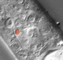
Pharyngeal Sensory Neuron I5
Description: I5 is a pharyngeal sensory neuron. Its cell body is in the ventral terminal bulb. It has processes through most of the terminal bulb, and processes in the subventral isthmus extend to the metacorpus, where they join the pharyngeal nerve ring.
When I5 is killed, pumps become slightly briefer than normal. This effect is probably caused by relief of M3 inhibition, since M3 acts to make pumps shorter than normal, and an I5-M3-worm has long pumps like M3-worms. Some I5-worms show a slight slippery isthmus defect. Growth is slightly slower than normal. (See Pharynx section; Albertson and Thomson, 1975; I5 neuron page.)
Pharynx Focal Plane 2
|
Identification: I5 is a large neuronal nucleus on the ventral midline of the terminal bulb. It is immediately anterior to mc3V, a big fried-egg nucleus, and posterior to the two side-by-side ventral g1 nuclei, also big fried eggs. |
|
|
