|
Pharynx Atlas Home
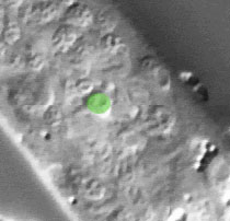
The pictures that comprise the Pharynx Atlas are a focal series through the terminal bulb of the
pharynx. The worm is lying on its right side, with anterior towards the upper left and posterior towards the lower right. Dorsal is the upper right and ventral is the lower left.
This is the first image and is a
superficial cut through the very leftmost edge of the pharynx.
Subsequent images go consecutively deeper, until the last goes through
the very rightmost edge. The focal planes are not equally spaced. They
were chosen so that all the nuclei in the terminal bulb would be
clearly visible in at least one image.
Click on individual pharyngeal cells to identify them.
Pharyngeal Terminal Bulb Muscle pm6VL
Description: The three pm6 muscle cells form most of the anterior half of the
terminal bulb. The cells are dorsal, left subventral, and right
subventral, and are separated from each other by the mc3 marginal
cell.
Pharynx Focal Plane 1
|
Identification: The pm6's are large fried eggs easily recognized by position and by
the line they form with pm7 and a neuron. pm6D is on the dorsal
midline, straight anterior from pm7D and just posterior to I4. pm6VL
and pm6VR are subventral, straight anterior of pm7VL and pm7VR, and
just posterior to the very small M2 neurons. |
|
|
