|
Pharynx Atlas Home
The pictures that comprise the Pharynx Atlas are a focal series through the terminal bulb of the
pharynx. The worm is lying on its right side, with anterior towards the upper left and posterior towards the lower right. Dorsal is the upper right and ventral is the lower left.
This is the first image and is a
superficial cut through the very leftmost edge of the pharynx.
Subsequent images go consecutively deeper, until the last goes through
the very rightmost edge. The focal planes are not equally spaced. They
were chosen so that all the nuclei in the terminal bulb would be
clearly visible in at least one image.
Click on individual pharyngeal cells to identify them.
Pharyngeal Motor Neuron M2L
Description: The two M2s are pharyngeal motor neurons of unknown function. Their
cell bodies are in the subventral anterior terminal bulb. They extend
processes anteriorly through the subventral isthmus to innervate
isthmus muscle (pm5) throughout its length. At the metacorpus the
processes enter the pharyngeal nerve ring and synapse on pm4. (See Pharynx section; Albertson and Thomson, 1975.)
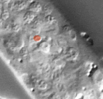
Pharynx Focal Plane 1
|
Identification: M2 is a very small nucleus. It is immediately anterior to pm7V, a big
fried-egg nucleus. The line of three cells pm6V (also a big
fried-egg), pm7V, and M2 extending from posterior to anterior along
the side of the terminal bulb is easy to recognize. |
|
