|
Cell Identification: Post Cloacal Sensilla and Spicule Nuclei
by L. Rene Garcia
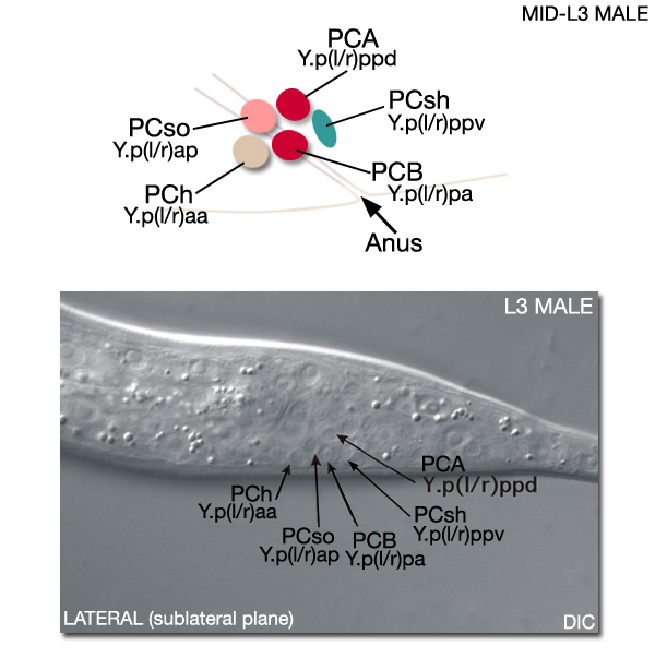
Mid-L3
Post cloacal cells from the Y.p lineage can be seen as early as the beginning of L3. These cells can be found flanking the left and right edges of the anus. They have a pattern of two cells on top of three cells on the bottom that persist up to mid L4. Usually Y.p(l/r)ap (PCso) and Y.p(l/r)aa (PCh) border the anal midline. Y.p(l/r)ppv (PCsh) is the most posterior, has a bean-shaped nucleus and is in a focal plane that is more internal and towards the anal space than the other cells. Again this persists through mid L4.
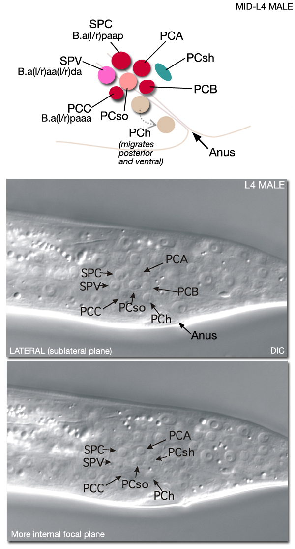
Mid L4
Mid L4, (when the tail spike is almost, but not completely retracted) is the best time to see all post cloacal cells as well as SPV and SPC (B lineage derived). Again these cells flank the anal region. The Y.p lineage cells keep their 2/3 cell pattern, put the pattern rotates anteriorly and PCh migrates ventrally. PCC (the sister of SPC) is anterior and ventral to PCso and sometimes this PCS (post cloacal sensilum) neuron is underneath SPV. PCB is slightly in an outward focal plane relative to PCso and PCA, whereas PCsh is in a slightly inward focal plane.
|
