|
REPRODUCTIVE SYSTEM OF THE MALE - SOMATIC GONAD
 Click pictures for new window with figure and legend Click pictures for new window with figure and legend
1 Male Somatic Gonad Development
1.1 Gonad Cell Generation
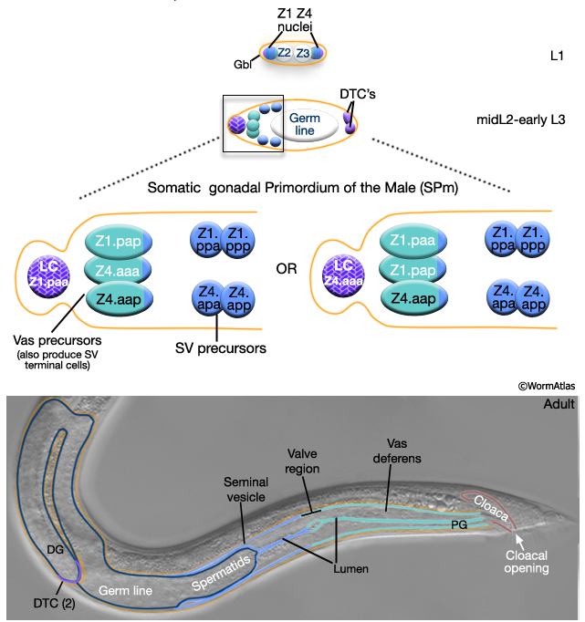
The male somatic gonad refers to the non-germline component of the gonad. It consist of 3 tissues: the distal tip cells (DTCs), the seminal vesicle (SV) and the vas deferens (Vas) (MaleReproFIG 2, Kimble and Hirsh, 1979). All cells of the somatic gonad develop from Z1 and Z4 of L1 gonad primordium. By L2, Z1/Z4 have generated 10 descendents: 2 DTCs, terminal somatic cells that will regulate germline patterning, 3 vas deferens and 4 seminal vesicle blast cell precursors and 1 linker cell (LC), a transient cell that functions in gonad elongation (Kimble and Hirsh, 1979). These cells organize to form the Somatic gonadal Primordium of the male (SPm) which establishes asymmetry of the future male gonad (MaleReproFIG 2). Between early-L3 and mid-L4 the SV and Vas precursors divide to generate a total of 53 progeny (23 SV cells and 30 Vas terminal cells). During this period, the primordium expands due to cell proliferation of the somatic and germ line lineages.
1.2 Gonad Elongation
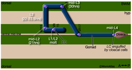
The gonad also elongates during this period guided by the male-specific linker cell (LC). The trajectory of its migration establishes the characteristic J-shape of the male gonad (MaleReproFIG 3; Hedgecock et al., 1987; Antebi et al, 1997). In hermaphrodites, this leader cell function is performed by the DTCs, which also regulate germ cell patterning. The male linker cell, however does not share this second function with the hermaphrodite DTCs and is required only for gonad migration. Possibly because of this functional difference, the male linker cell and DTCs have different morphologies. The linker cell is rhomboid shaped and does not extend cytonemes (tendril-like processes) proximally over the germ line (MaleReproFIG 4).
MaleReproFIG 3: Migration of the male linker cell. Diagram of a worm opened along the dorsal midline. Landmarks (VR15 etc.) are indicated. (BWM) Body wall muscle; (hyp) hypodermis. (Adapted from Hedgecock et al., 1987; Antebi et al., 1997.)
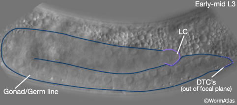
MaleReproFIG 4: The male linker cell. Nomarski DIC of male gonad featuring the linker cell (LC) and the distal tip cell (DTC) during early-mid L3 stage, lateral view.
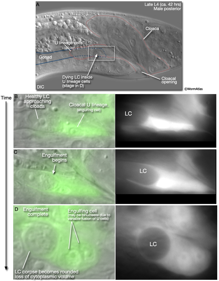
Progression of gonad elongation can be used to roughly estimate male larval stage. Towards the end of its migration the linker cell passes between U lineage cells of the developing cloaca (MaleReproFIG 5). One of these cells (U.lp or U.rp, or their fusion products) engulfs the linker cell. This opens the passageway between the lumens of the vas deferens and the cloaca (for more about U cells see Male Reproductive System - Proctodeum).
MaleReproFIG 5: Engulfment of the male linker cell by cloacal cells. A. Nomarski DIC image of the male posterior region of a late L4 animal. Lateral view of the cloacal cells engulfing the male linker cell (LC). B-D shows the engulfment process with time. Epifluorescent images from transgenic animals expressing the lin-48::GFP reporter gene. (Strain source: A.D. Johnson and H. Chamberlin.) (Image source: M. Abraham and S. Shaham.)
2 The Distal Tip Cells
In males, the two distal tip cells (DTC's) are found together at the distal end of the gonad (MaleReproFIG 6). This arrangement is established early in gonad development during formation of the SPm (MaleReproFIG 2; Kimble and Hirsh, 1979). As in the hermaphrodite the DTCs in the male are required to maintain the nearby germ cells in a mitotic state or to prevent them from entering meiosis (for more on meiotic regulation by the DTC see Hermaphrodite Handbook - Somatic Gonad; Kimble and White, 1981). In contrast to the hermaphrodite, however, the male DTCs do not migrate during gonad development; this function is fulfilled by the male linker cell (see above MaleReproFIG 3, 4 and 5).
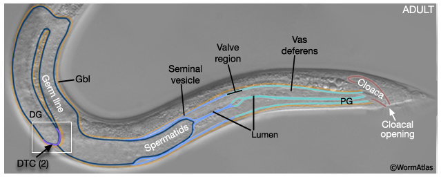
MaleReproFIG 6: The distal tip cells. Nomarski DIC image of the distal end of the adult male gonad, lateral view. (DG) Distal gonad; (PG) proximal gonad; (Gbl) gonadal basal lamina; (SV) seminal vesicle; (Vas) vas deferens; (DTC) distal tip cell.
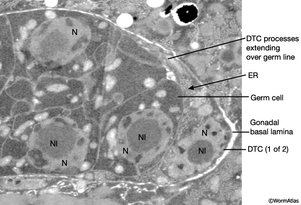
Superficially male DTCs appear to have a similar morphology to those of the hermaphrodite (for a detailed description of hermaphrodite DTCs see Hermaphrodite Handbook - Somatic Gonad). The DTC is a single, large somatic cell that has a large nucleus located at its distal edge. The cell forms a close-fitting cap over the most distal 6-10 germ cells (MaleReproFIG 7). No intervening basal lamina or specialized intercellular junctions are found between the DTC and germ cells.
MaleReproFIG 7: Ultrastructure of the male distal tip cell. TEM of the distal tip cell (DTC) as it forms a cap over the most distal germ cells, longitudinal view. (Image source: him5-25 [Hall] 2307.) (N) Nucleus; (NI) nucleolus; (ER) endoplasmic reticulum. |
3 The Seminal Vesicle
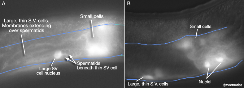
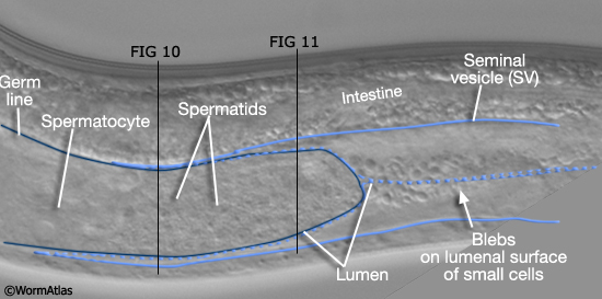
The seminal vesicle (SV) consists of an inner tube of 20 apparent secretory cells that are surrounded by a thin sheet of the cytoplasmic processes formed by 3 large cells. Shortly after their differentiation the small cells have a granular appearance when viewed with DIC optics and small blebs are apparent on their lumenal surfaces (MaleReproFIG 8; Kimble and Hirsh, 1979). The seminal vesicle has a larger outer diameter than either the testis or vas deferens. The small cells derived from the four seminal vesicle precursors of the SPm while the large, thin cells are the most distal daughters of the vas deferens precursors (MaleReproFIG 2). These large cells are born proximal to the seminal vesicle and then enlarge, flatten and spread over smaller cells encapsulating them and germline cells. Their thin, sheet-like morphology and extension over the germ line is reminiscent of the distal sheath cells of the hermaphrodite gonad. (MaleReproFIG 9, 10 and 11).
MaleReproFIG 8: The male seminal vesicle. Nomarski DIC picture of a young adult male, lateral view, medial plane.
MaleReproFIG 9: Seminal vesicle cells. Epifluorescent images from transgenic animals expressing the K09C8::GFP reporter gene, lateral view, lateral plane. (Strain source: K. Theomke, W. Chang and D. Zarkower.) (Image source: M. Abraham and S. Shaham.) B image is a lower exposure of animal in A.
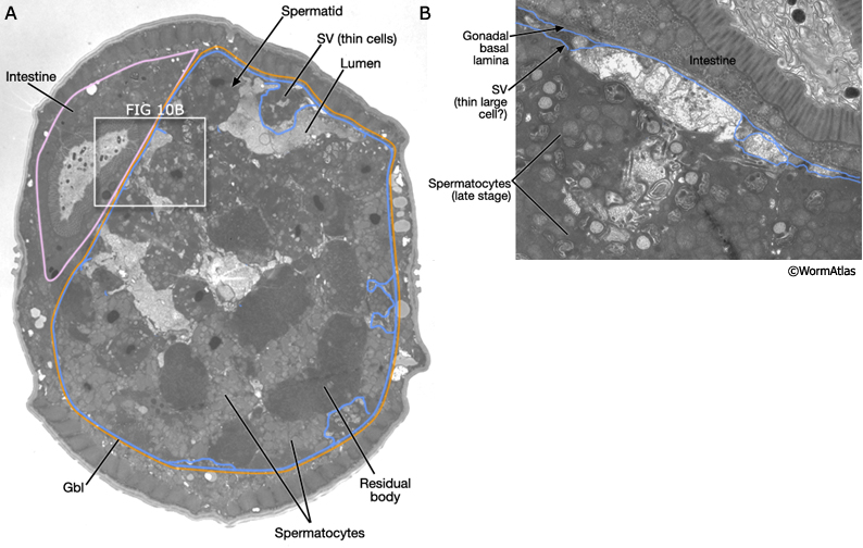
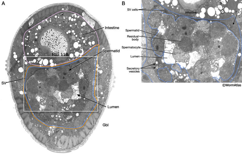
The seminal vesicle stores spermatids until ejaculation. Spermatids are the sessile precursors to amoeboid mature spermatozoa. Spermatids are formed by the process of spermatogenesis where spermatocytes detach from the rachis of the proximal germ line and undergo 2 meiotic divisions (MaleReproFIG 8, 10 and 11; Wolf et al., 1978; see Male Reproductive System - Germ LIne).
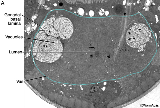
MaleReproFIG 10: Ultrastructure of the seminal vesicle. A. Low-power TEM highlighting the seminal vesicle and adjacent gonadal basal lamina, transverse view. (Image source: him5-25 [Hall] 1304.) B. Higher magnification TEM of white boxed region in A, transverse view. (Image source: him5-25 [Hall] 1593.) (Gbl) Gonadal basal lamina; (SV) seminal vesicle.
MaleReproFIG 11: Ultrastructure of the seminal vesicle. A. Low-power TEM highlighting the seminal vesicle and adjacent gonadal basal lamina, transverse view. (Image source: him5-25 [Hall] 1226.) B. Higher magnification TEM of white boxed region in A, transverse view. (Image source: him5-25 [Hall] 1226.) (Gbl) Gonadal basal lamina; (SV) seminal vesicle.
4 The Vas Deferens
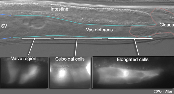
The vas deferens (vas) is a complex secretory tube that extends from the seminal vesicle to the cloaca. It is composed of 30 cells that can be divided into at least 3 cell types based on morphological differences in secretory granules alone (N. Wolf, J. Kimble and D. Hirsh unpublished observations). The anterior end of the vas appears to act as a valve that may regulate sperm release from the seminal vesicle during ejaculation (MaleReproFIG 12 and 13). The anatomical characteristics of valve cells are not well defined. However, Gower et al., 2005 have identified a potential molecular marker, the itr-1 reporter gene, which exhibits elevated expression in cells of this region (MaleReproFIG 12). Differences in cell shape are also apparent along the length of the vas. Posterior to the valve, some cells are cuboid in shape while closer to the cloaca, vas cells are elongated (MaleReproFIG 12).
MaleReproFIG 12: Regions of the vas deferens. Top DIC image of the male posterior region highlighting the vas deferens. Bottom Epifluorescent images from transgenic animal expressing the itr-1::YFP reporter gene featuring the differently shaped cells in the 3 different regions of the vas deferens. (Strain source: G. Schindelmann and P.W. Sternberg.)
|
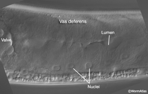
MaleReproFIG 13: The vas deferens. Nomarski DIC image of the vas deferens of an adult male, ventral view.
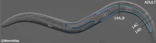
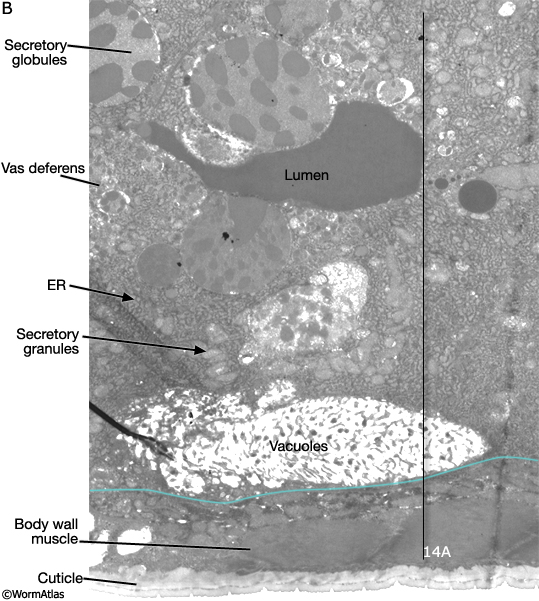
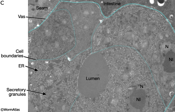
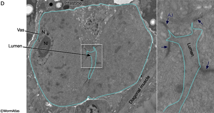
Consistent with a secretory function, cells of the vas deferens are rich in ribosomes, ER,secretory granules and potential storage vacuoles (MaleReproFIG 14, 14A, 14B, 14C and 14D).
MaleReproFIG 14: Adult male gonad. Low-power Nomarski DIC of an adult male with the seminal vesicle (SV) and vas deferens (Vas) highlighted, left lateral view. Black lines indicate regions with EM images shown in A-D, below.
MaleReproFIG 14A: Ultrastructure of anterior vas deferens. TEM of region indicated by black line in FIG 14, transverse view. (Image source: him5-25 [Hall] Z059.)
MaleReproFIG 14B: Ultrastructure of anterior vas deferens. TEM of region indicated by black line in FIG 14, longitudinal view. Cell boundaries are highlighted by dotted blue lines. (Image source: him5-25 [Hall] 1633.)
MaleReproFIG 14C: Ultrastructure of posterior vas deferens. TEM of region indicated by black line in FIG 14, transverse view. (Image source: him5-25 [Hall] 1022.) (N) Nucleus; (NI) nucleolus; (ER) endoplasmic reticulum.
MaleReproFIG 14D: Ultrastructure of posterior vas deferens. TEM of region indicated by black line in FIG 14, transverse view. (Image source: him5-25 [Hall] Z854.) Panel on right shows detail of the region around with lumen with arrows indicating the location of the adherens junctions (AJ).
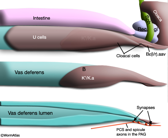
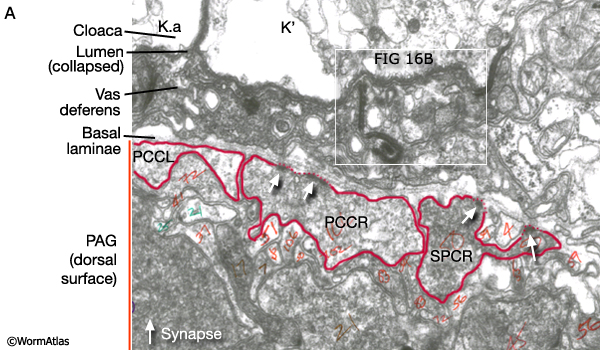
The most proximal, ventral cell of the vas and its neighbor, cloacal cell Bε(l/r).aav, are innervated by neurons of post-cloacal sensilla (PCBL/R and PCCL/R) and spicule neurons (SPCL/R) (MaleReproFIG 15 and 16A; Male Wiring Project). Synapses occur where axons run in the dorsal PAG (pre-anal ganglion), directly beneath these gonadal cells. The post-cloacal sensilla and spicule function in the steps that precede ejaculation, namely vulval location and spicule insertion (see Mating Behavior Movie in Introduction to Male Anatomy ). Direct gonadal innervation by SPCL/R, PCBL/R and PCCL/R suggests that vulval contact and fully extended spicules may trigger this step (reviewed in WormBook: Male Mating Behavior - Barr and Garcia).
MaleReproFIG 15: Cloacal-gonad connection. Diagram of an adult male tail, lateral view. Based on the N2Y (MRC) LP EM series. (Rec. gl) Rectal gland: (PAG) pre-anal ganglion; (PCS) post-cloacal sensilla; (Sph) sphincter muscle.
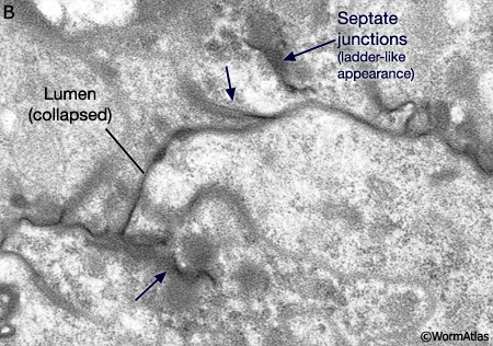
The innervated posterior vas cell and Bε(l/r).aav contain numerous electron-dense vesicles that may be secretory (MaleReproFIG 16A). These cells line the vas and cloacal lumens, respectively. Their lumenal (apical) surfaces are extensively folded and bear septate junctions (MaleReproFIG 16A and 16B). This is reminiscent of the hermaphrodite spermatheca in hermaphrodites where the lumen may close and open by unzipping of septate junctions. Possibly opening and closing of the vas/cloacal lumen may be regulated in response to inputs from the dorsal PAG.
MaleReproFIG 16A: Innervation of the vas deferens. TEM of cells lining the lumen of the vas deferens shows electron dense vesicles suggesting a secretory function, transverse view. (Image source: N2Y PAG [MRC] 875.) (PAG) pre-anal ganglion.
MaleReproFIG 16B: Septate junction in the vas deferens. High power EM of region indicated by white box in 16A, transverse view. Arrows point to septate junctions. (Image source: him5-25 [Hall] 4656.)
5 List of Male Somatic Gonad Cells (See male Z lineage)
DTC (gon male dtc) :
Z1.a
Z4.p
Seminal vesicles (gon male sves)
Outer cells
Z1.pappp
Z1.paapp or Z4.aaapp
Z4.aappp
Inner cells
Z1.pp (x10)
Z4.ap (x10)
Vas deferens (gon male vdef)
Z1.pap (x10)
Z4.aa (x20)
or
Z4.pa (x20)
Z4.aap (x10)
Linker cell
Z1.paa or Z4.aaa
6 References
Antebi, A., Norris, C.R., Hedgecock, E.M. and Garriga, G. 1997. Cell and Growth Cone Migrations. In C. elegans II (ed. D.L. Riddle et al.). Chap. 21. pp. 583-609. Cold Spring Harbor Laboratory Press, Cold Spring Harbor, New York. Article
Gower, N.J., Walker, D.S. and Baylis, H.A. 2005. Inositol 1,4,5-trisphosphate signaling regulates mating behavior in Caenorhabditis elegans males. Mol. Biol. Cell 16: 3978-86. Article
Hedgecock, E.M., Culotti, J.G., Hall, D.H. and Stern, B.D. 1987. Genetics of cell and axon migrations in Caenorhabditis elegans. Development 100: 365-382. Article
Kimble, J. and Hirsh, D. 1979. The postembryonic cell lineages of the hermaphrodite and male gonads in Caenorhabditis elegans. Dev. Biol. 70: 396-417. Article
Kimble, J. and White, J.G. 1981. On the control of germ cell development in Caenorhabditis elegans. Dev. Biol. 81: 208-19. Abstract
Miskowski, J., Li, Y. and Kimble, J. 2001. The sys-1 gene and sexual dimorphism during gonadogenesis in Caenorhabditis elegans. Dev. Biol. 230: 61-73. Article
Wolf, N., Hirsh, D. and McIntosh, J.R. 1978. Spermatogenesis in males of the free-living nematode, Caenorhabditis elegans. J. Ultrastruct. Res. 63: 155-69. Abstract
|

This chapter should be cited as: Lints, R. and Hall, D.H. 2009. Male reproductive system, somatic gonad. In WormAtlas. doi:10.3908/wormatlas.2.15
Edited for the web by Laura A. Herndon. Last revision: July 22, 2013. |
|