|
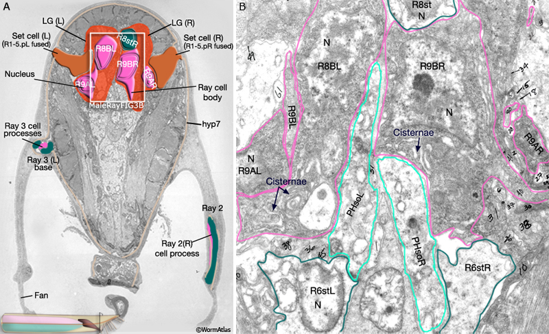
MaleRayFIG 3: Ultrastructure of the lumbar ganglia.
A. Low-power TEM highlighting the lumbar region with the rays, transverse section. (Image source: N2Y [MRC] 27.)
B. Higher magnification TEM of the ray cell bodies within the lumbar ganglia from boxed region of A, transverse view. (N) Nucleus; (PhsolL/R) Phasmid socket cells. (Image source: N2Y [MRC] 1350.) |