|
RectFIG 3A-E: The rectum.
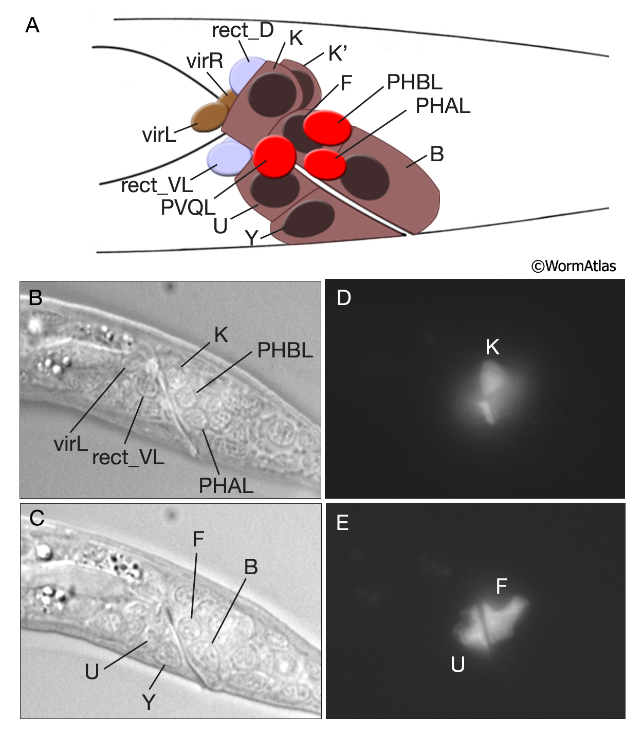
A. Graphic rendition of rectal cells in early L1 larva. Their nuclei are shown as dark ovals. Nuclei of some of the neurons in this region, the gland cells and the valve cells are also shown. The rectal epithelial K and K’ cells lie as a left and right pair, whereas the remaining rectal epithelial cells, B, F, U and Y are placed dorso-ventrally around the lumen. (Based on Sulston et al, 1983).
B-E. Rectal epithelial cells in L1 larva. B & C DIC images, left lateral views. B is taken from a plane more lateral than C and shows the nucleus of K as well as the neuron nuclei. Four other rectal nuclei are seen at a medial plane in C. D & E Epifluorescent images from the same animal expressing the transgene lin-48::GFP in some rectal epithelial cells. (Strain Source: H. Chamberlin.) Magnification, 600x.
See also RectFIG 3F and 3G.
Click on picture for full resolution image.
|