|
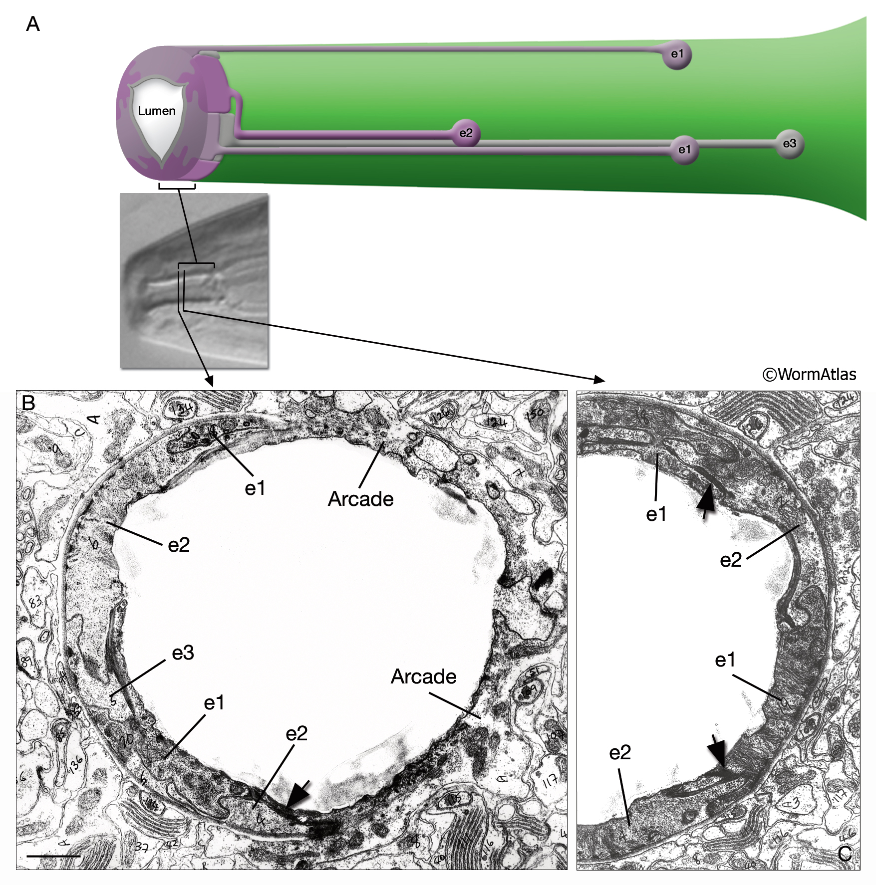
PhaFIG 5A-C: Pharyngeal epithelium.
All except insets are transverse section TEM images.
A. Ring of nine epithelial cells encircle the anterior of the pharynx. The dorsal and subventral sides of this circle are occupied by the e1 cells. Three e2 cells are localized at the corners of the lumen. Their thin processes make a short clockwise turn in front of pm1 to connect to the buccal cavity portion. Posterior to e1, three e3 cells occupy the dorsal and subventral regions of the posterior buccal cavity. The cell bodies of e1, e2 and e3 are located, respectively, about 30, 20 and 35 μm posteriorly within the corresponding nerve cords. (Based on Albertson and Thomson, 1976.)
(Inset) DIC image indicating the levels of B and C.
B. Transition between the posterior arcade cells and the pharyngeal epithelium. The cells are connected to each other by large adherens junctions (arrow), sealing the pharynx to the buccal tissues. Numbers seen in B and in C-E are from the original prints. Bar, 1 μm. (Image source: N2T [MRC] A60-8.)
C. Relationship of e1 and e2 cells at the anterior of the pharyngeal epithelium. Cells within each set do not touch to each other, but e1 and e2 cells are connected to each other by adherens junctions (arrows). (Image source: N2T [MRC] A60-22.)
See also PhaFIG 5D&E.
Click on picture for full resolution image.
|