|
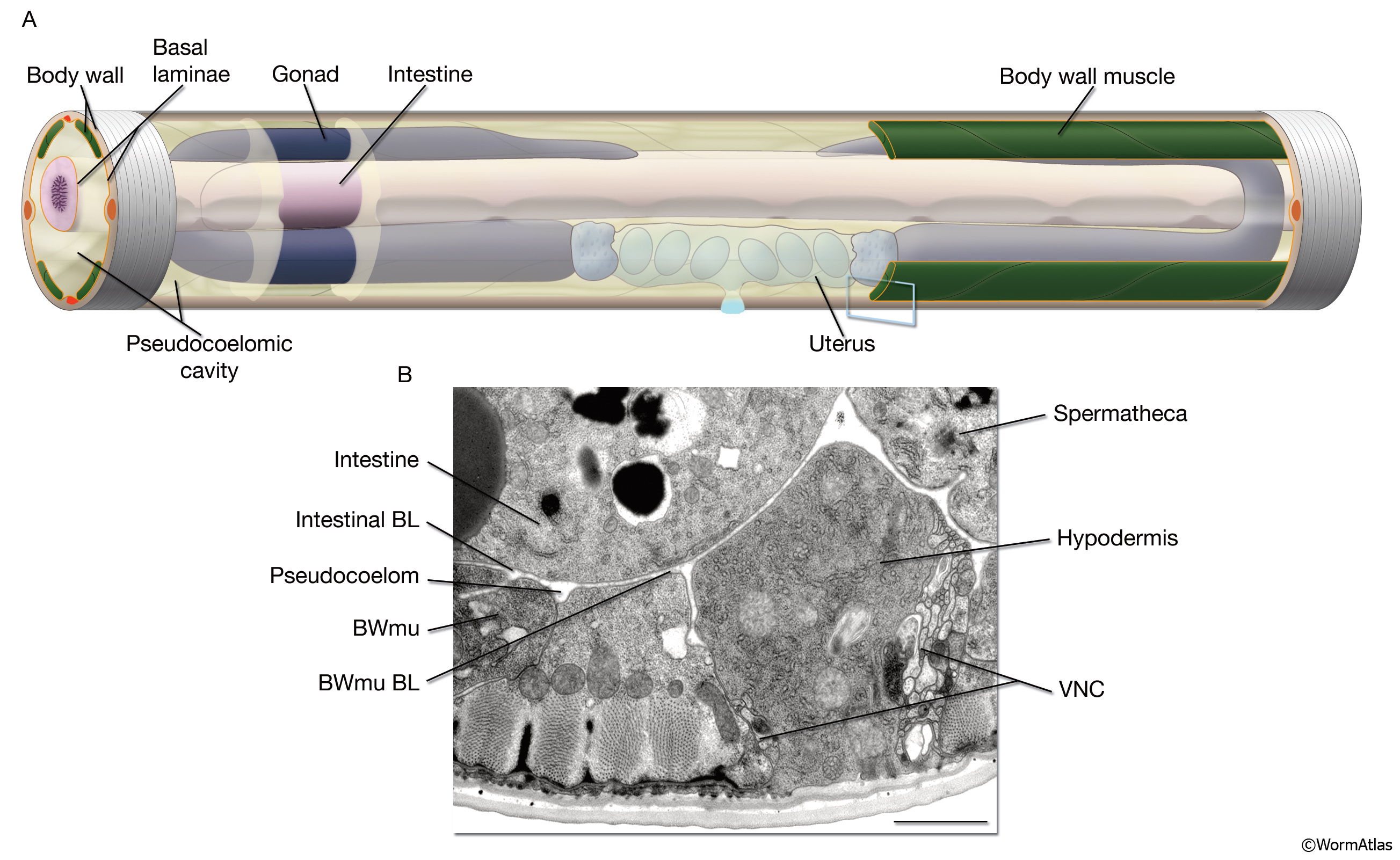
PeriFIG 1: Pseudocoelom (body cavity).
A. Graphic representation of the body cavity and pseudocoelomic fluid. To emphasize its space filling character, the pseudocoelom in this stylized image is shown larger than its actual volume. Head and tail regions not shown. Most of the left side body wall, except some anterior and posterior hypodermis and posterior muscle quadrants as well as a small anterior portion of the pseudocoelom, are removed to display the relation between the pseudocoelom and internal tissues. Pseudocoelomic fluid bathes all tissues. The pseudocoelom is lined by basal laminae (BL) (orange lines).
B. The body cavity (pseudocoelom) of C. elegans is lined by the BL of the body wall and internal tissues, rather than mesodermal cells, as would be seen in a true coelomic cavity. Transverse thin section corresponding to blue box in A. (Bwmu) Bodywall muscle; (VNC) ventral nerve cord. Bar, 1μm. (Image source: [Hall] N483.)
Click on picture for full resolution image.
|