|
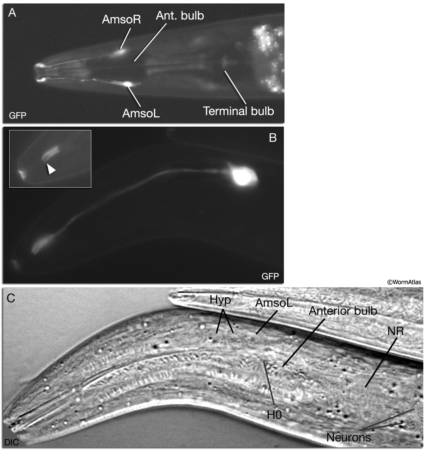
NeuroFIG 28: Structure of the amphid socket cell.
A. Epifluorescent image from an animal expressing the itr-1pB::GFP reporter gene, dorsal view. Amphid socket cells are located on each side of the anterior bulb, close to the sensilla. Magnification, 400x. (Strain source: H. Baylis and N. Gower.)
B. Epifluorescent image from an animal expressing the T16G1.8::GFP reporter gene, left lateral view. Amphid socket cells have simple morphologies with a small cell body, a thin process, and a small cylindrical ending (arrowhead in inset) on the lateral sides of the lips. Magnification, 600x. (Strain source: The Genome BC C. elegans gene expression consortium [McKay et al. 2004].)
C. DIC, same animal as in B. Amphid socket nucleus has an epithelial appearance. It is located close to the anterior lateral hypodermal and first seam nuclei.
Click on picture for full resolution image.
|