|
NeuroFIG 26: Amphid neurons and amphid nerve.
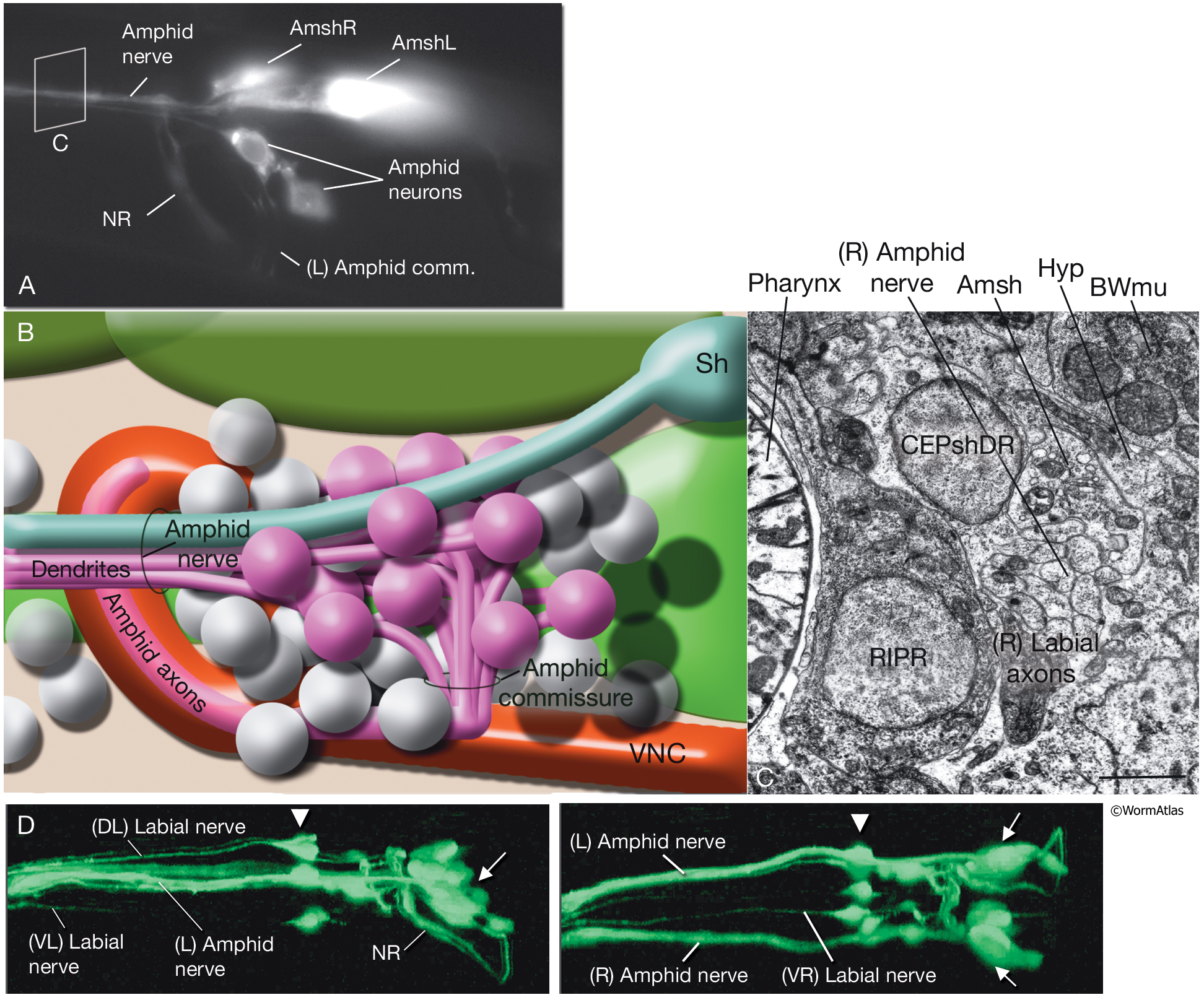
A. Epifluorescent image, left lateral view. Each of the bilaterally symmetric amphid nerves contains dendrites of 12 amphid neurons and the process of one amphid sheath cell. The amphid nerves travel on the lateral sides of the pharynx to the lips. The axons of the amphid neurons enter the amphid commissures on each side to reach the VNC and NR. DiI-stained animal expressing ver1::GFP marker in amphid sheath cells. Magnification, 400x. (Strain source: R. Roubin, C. Popovici, and S. Shaham.)
B. Illustration of left-side amphid neurons, amphid commissure, and amphid nerve. Amphid neuron cell bodies are located posteriorly to the nerve ring, whereas amphid sheath cells are on the dorsolateral sides of the terminal bulb of the pharynx.
C. TEM of right lateral region, anterior to the NR along the pharyngeal isthmus, transverse section. The amphid sheath process and the amphid neuron dendrites constitute the amphid nerve bundle, which at this level is located next to the posteriorly extending axons of the lateral labial neurons. The amphid nerve and the labial axons are sandwiched between cell bodies of NR neurons and the pharynx on the inside and the hypodermis and muscle on the outside. Bar, 1 μm. (Image source: N2U [MRC] A176-7.)
D. Epifluorescent images of amphid (arrows) and IL1 (arrowheads) neurons. (Left panel) Left lateral view; (right panel) ventral view. Strain marker: Y105E8A.5::GFP. (Image source: R. Newbury. The Genome BC C. elegans gene expression consortium [McKay et al., 2004].)
Click on picture for full resolution image.
|