|
NeuroFIG 9A-C: Mechanosensory neurons that sense gentle touch.
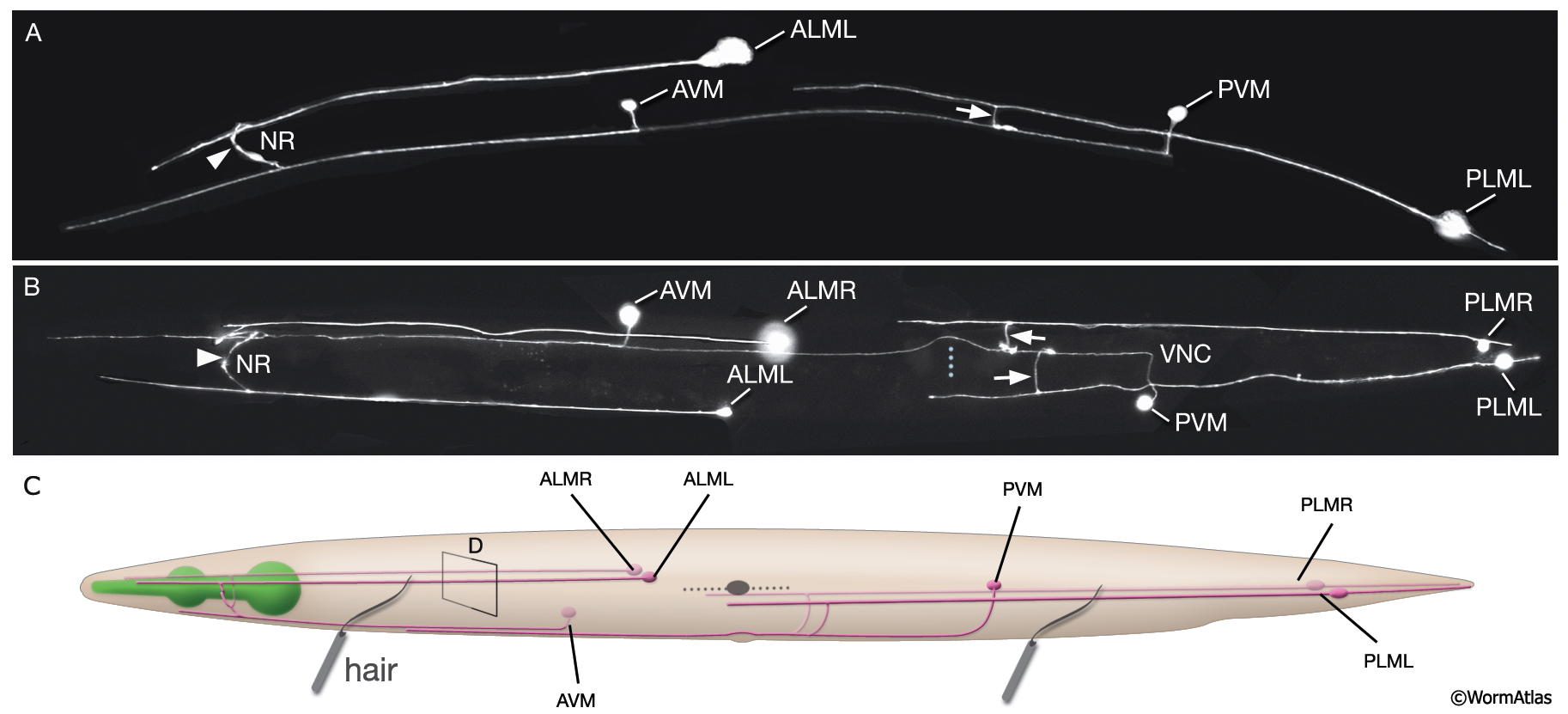
A&B. Fluorescent micrographs of an adult animal expressing the reporter gene mec-4::GFP in touch receptor neurons. A. Left lateral view. B. Ventral image taken from the dorsal side. The unpaired AVM and PVM are located sublaterally on the right anterior ventral side and left posterior ventral side of the animal, respectively. Their processes enter the VNC via commissures and run in an extreme ventral position near the cuticle within the VNC. The PVM process ends in the anterior body. The AVM process terminates slightly posterior to the anterior bulb of the pharynx; however, at about 1–2 μm anterior to the excretory pore, it sends off branches that enter the NR on both sides. The ALM cell bodies are located laterally in the midbody where PLM processes end. The process of each ALM runs near the dorsal edge of the lateral hypodermal ridge on each side, in close association with the process of ALN. ALMprocesses send off branches about 10 μm anterior to the excretory pore. These processes enter the NR subdorsally, end shortly after they meet, and make gap junctions with the branches of the AVM (arrowheads). ALM and AVM make synapses with command interneurons within the NR. The PLM cell bodies are situated in the lumbar ganglia; their anterior processes run near the ventral edge of the lateral hypodermal ridges, in close association with the processes of PLN. The PLM processes turn ventrally and enter the VNC near the vulva (arrows). VNC branches terminate after a short distance, whereas the main process terminates within midbody region, adjacent to where ALM is located. (NR) Nerve ring; (blue dotted line) position of vulva. Magnification, 400x. (Strain source: M. Driscoll.) C. Graphic rendition of touch receptor neurons as seen from the left side. Stroking the animal’s body with an eyelash generates a gentle touch stimulus that stimulates the touch receptor neurons and initiates an avoidance response. (Gray dotted line) Lateral midline; (gray oval) position of the CAN neuron, depicted for position comparison.
See also NeuroFIG 9D-J
Click on picture for full resolution image.
|