|
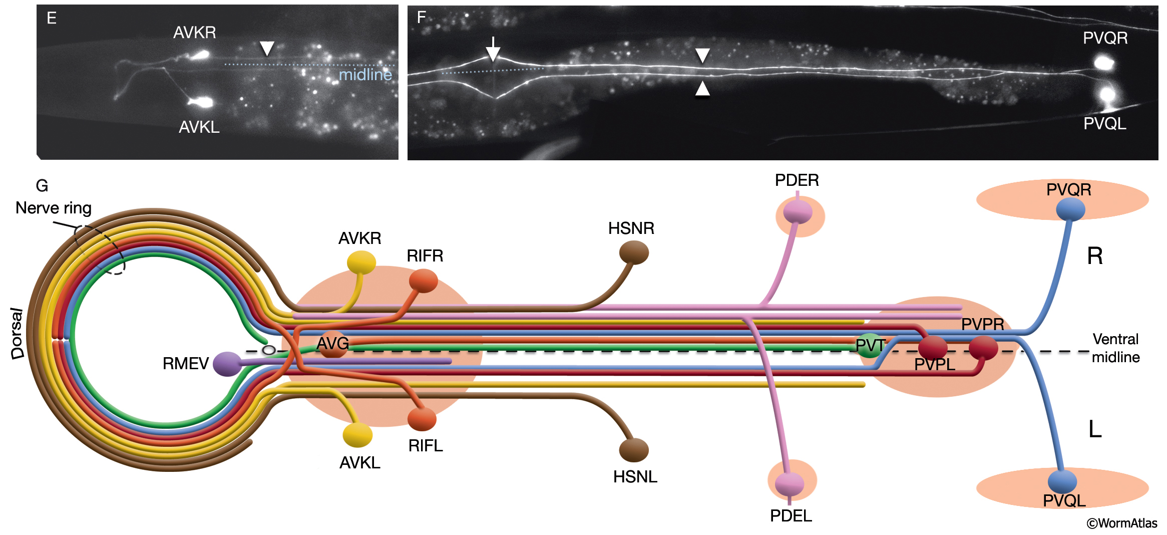
NeuroFIG 4E-G: Asymmetries in the C. elegans nervous system.
E. Ventral view seen from the dorsal. The AVKR process does not cross to the right side after exiting the NR on the left and remains on the left side of the VNC (arrowhead). (Blue dotted line) Ventral midline. Epifluorescent image of a transgenic animal expressing the flp-1::GFP transgene in AVK neurons. Magnification, 600x. (Strain source: B. Wightman and C. Li.)
F. Ventral view seen from the dorsal. PVQ neurons grow processes in the left and right tracts of the VNC (arrowheads). After entering the preanal region from the left lumbar commissure, the PVQL process briefly runs along the process of PVQR before extending into the left tract of the VNC. (Arrow) Vulva; (blue dotted line) midline. Epifluorescent image of a transgenic animal expressing the sra-6::GFP transgene in PVQ neurons. Magnification, 600x. (Strain source: T. Sarafi-Reinach and P. Sengupta.)
G. Schematic view of neurons that pioneer the left and right fascicles of the VNC. The body posterior to the NR is shown as if opened along the dorsal midline in cylindrical projection while the nerve ring is flattened toward the anterior. (Circle) Excretory pore at the anterior of the ventral midline; (light red ovals) ganglia. For clarity, neurons are given individual colors that are different from the color code used throughout this Atlas. (Based on White et al., 1986; Wadsworth et al., 1996.)
See also NeuroFIG 4A-D
Click on picture for full resolution image.
|