|
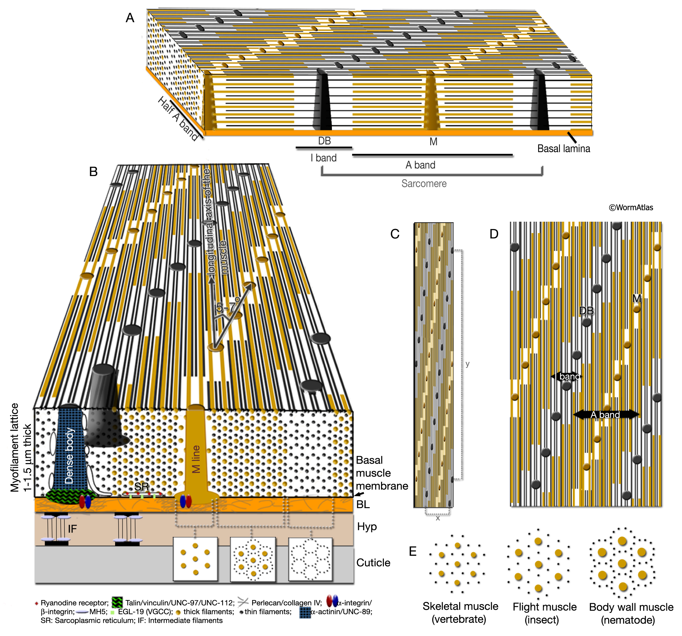
MusFIG 9: Organization of the myofilament lattice.
Yellow lines) Thick filaments; (black lines) thin filaments; (black dots) DB; (brown dots) M lines.
A. Three-dimensional rendering of myofilament lattice as well as structure of the sarcomere.
B. Schematic illustration of a cross section of the myofilament lattice. In the muscle cell, the sheet of filaments lies inside the muscle membrane, which is separated from the underlying hypodermis by a 20-nm basal lamina (BL). Cuticle, in turn, lies outside the hypodermis (Hyp). The striations created by the thick and thin filaments are at an angle of 5–7° to the longitudinal axes of the filaments and the muscle cell (shown at an increased angle for emphasis; compare to C).
C. Accurate surface view of myofilament structure and filament packing. Note that the distance between DBs (black dots) on the longitudinal axis is more than 10 µm (y), whereas in the tranverse plane they are only about 1 µm apart (x), for a ratio of about 10:1 (R.H. Waterston, pers. comm.).
D. The surface view is laterally extended to better indicate the offset of contractile units (offset is increased for emphasis; compare with C).
E. Comparison of packing of thick (yellow dots) and thin (black dots) filaments at transverse section among the vertebrate skeletal muscle, the insect flight muscle, and the nematode body wall muscle.
Click on picture for full resolution image.
|