|
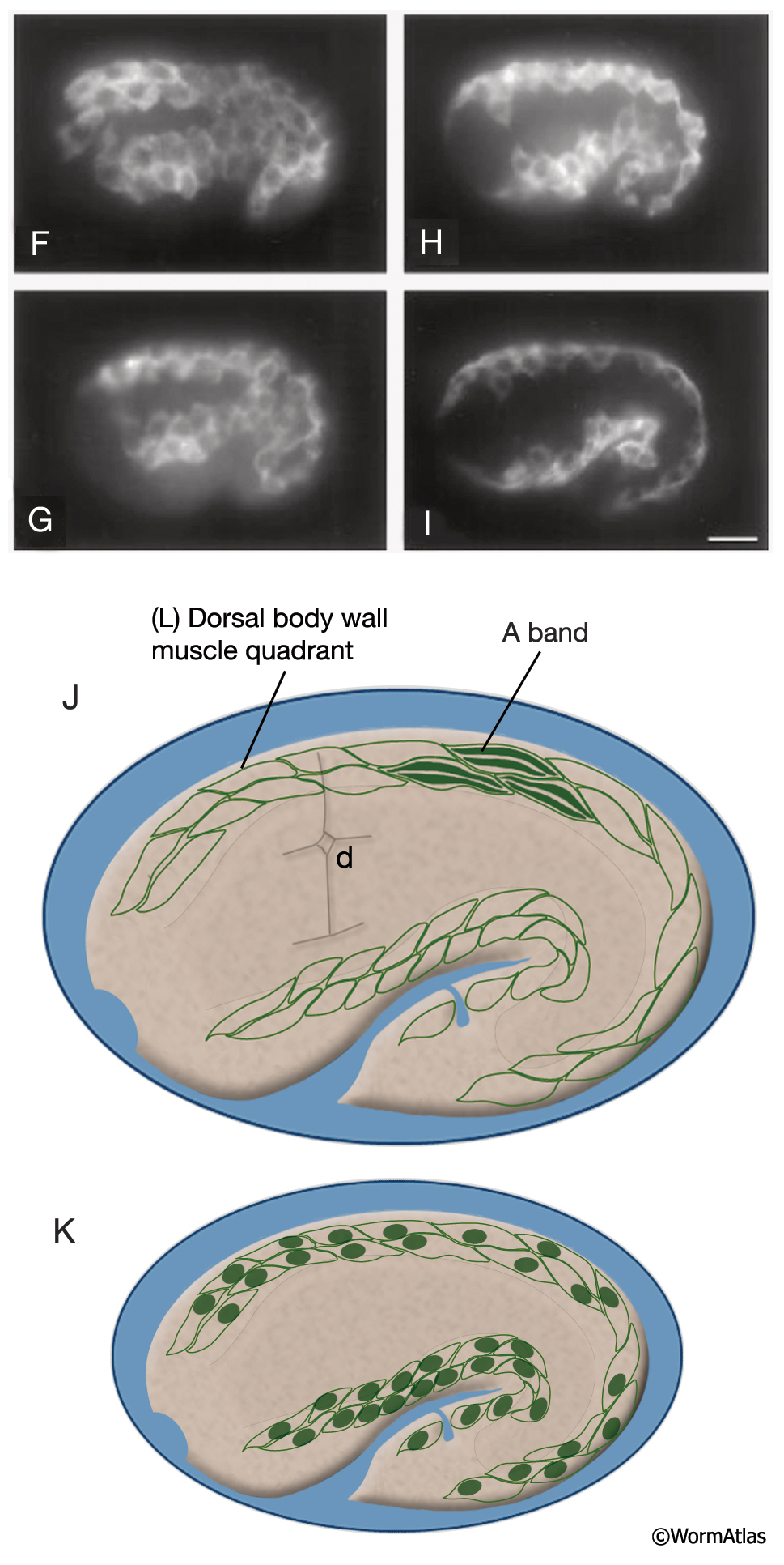
MusFIG 14F-K: Development of somatic muscle.
F-I. Developing muscles in embryos stained with the muscle marker NE8 4C6.3, left lateral view. Bar, 10 μm. (Reprinted, with permission, from Moerman et al., 1996. ©Elsevier.)
F. Approximately 310-minute embryo after first cell cleavage. Muscle cells start to migrate from a lateral position adjacent to seam cells to ventral and dorsal sides. The anterior part has separated, whereas the posterior half is still in a continuous sheet.
G. Approximately 330-minute embryo. The more posterior muscle cells also started to move dorsally and ventrally.
H. Approximately 350-minute embryo. Dorsal and ventral muscle quadrants have formed.
I. A 420-minute embryo (1.5-fold stage). Muscle cells become more flattened as myofilaments are forming.
J. Location of body wall muscle cells (green lines) in strips in the dorsal and ventral quadrants in a 1.75-fold (420–450 min) embryo just after acquisition of contractile function (compare to MusFIG 15), left lateral view. Arrangement of A bands is shown inside three muscle cells. There are four A bands across the quadrant because each muscle cell is only two sarcomeres wide. The position of the deirid (d) is also shown for relative position. (Based on Williams and Waterston, 1994; Moerman and Fire, 1997.)
K. Illustration of the relative positions of 40 body wall muscle nuclei on the left side of the embryo. (Based on Sulston et al., 1983.)
See also MusFIG 14A-E.
Click on picture for full resolution image.
|