|
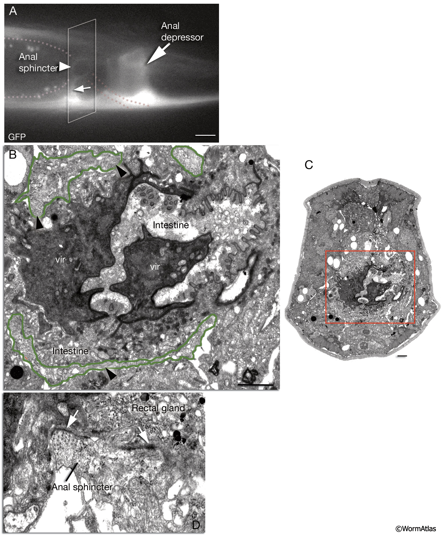
MusFIG 20: Anal sphincter muscle.
A. The anal sphincter is located at the junction of the posterior intestine and the rectum (dotted lines). It includes a toroidal part that encircles the posterior end of the intestine and four thin processes that attach to the dorsal and ventral body wall (small arrow [ventral left process] arrowhead [toroidal section]). (White rectangle) Level of the section in B and C. Epifluorescent image from a transgenic animal expressing the reporter gene unc-68::GFP, left lateral view. Bar, 10 μm. (Strain source: E. Maryon.)
B&C. Cross section of the anal sphincter muscle, TEM image. B. High power. C. Low power. (Arrowheads) Circularly oriented contractile elements within the toroidal section (outlined in green in B). Many of these myofibrils have no obvious attachments, but some indent deeply into the rectal gland tissue inside a few short processes. (vir) Intestinal-rectal valve. The level of the section is indicated in C. Bar, 1 μm. (Image source: JSE [MRC] 617-14.)
D. Short, medially directed processes extend from the toroid. These contain filaments that show electron-dense attachments (white arrows) to the rectal gland cells near the intestinal-rectal valve, TEM image, transverse section. (Image source: JSE [MRC] 617-119.)
Click on picture for full resolution image.
|