|
MusFIG 17: Pharyngeal muscles have single sarcomeres.
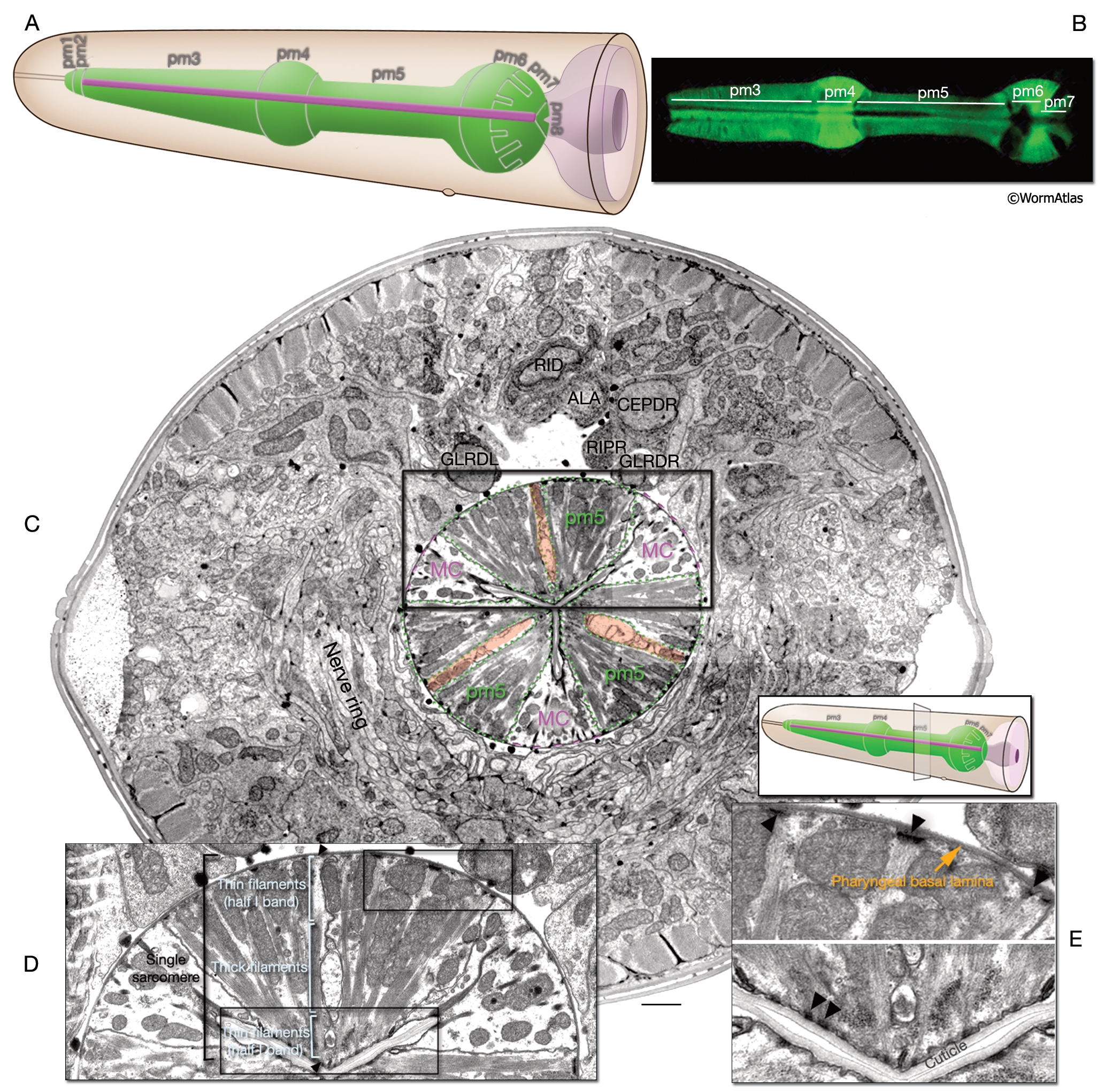
A. Schematic diagram of the pharyngeal muscles, which are organized into eight cell groups, pm1–pm8 (also called m1–m8) (Moerman and Fire, 1997). Indicated are the approximate location and shape of individual cells, as seen from the left lateral side. (Purple) Pharyngeal MC cells.
B. Epifluorescent image of pharyngeal muscles pm3–pm7 taken from a transgenic animal expressing the reporter gene C32F10.8::GFP. Muscles occupy positions within the pharynx as indicated. pm1, pm2, and pm8 do not express this marker. (Image source: R. Newbury. The Genome BC C. elegans gene expression consortium [McKay et al., 2004].)
C. Ultrastructure of single sarcomeres of pharyngeal muscle. TEM image of a cross section of the pharyngeal isthmus from a level at the posterior of the nerve ring (small graphic inset, right). pm5 muscle cells, MC cells, and nerve cords (red) show threefold symmetry within the pharynx. Bar, 1 μm. (Image source: N2U [MRC] A194 8-11.)
D. Each sarcomere of the pharyngeal muscle covers the radial length of the muscle cell, ending in a half I band (thin filaments) and electron-dense attachments (arrowheads) at each end. Thick filaments occupy the middle of each sarcomere.
E. On the pseudocoelomic face, each sarcomere ends in an electron-dense attachment (arrowheads) to the pharyngeal basal lamina. On the lumenal side, half I bands attach to the cuticle through electron-dense attachments (arrowheads).
Click on picture for full resolution image.
|