|
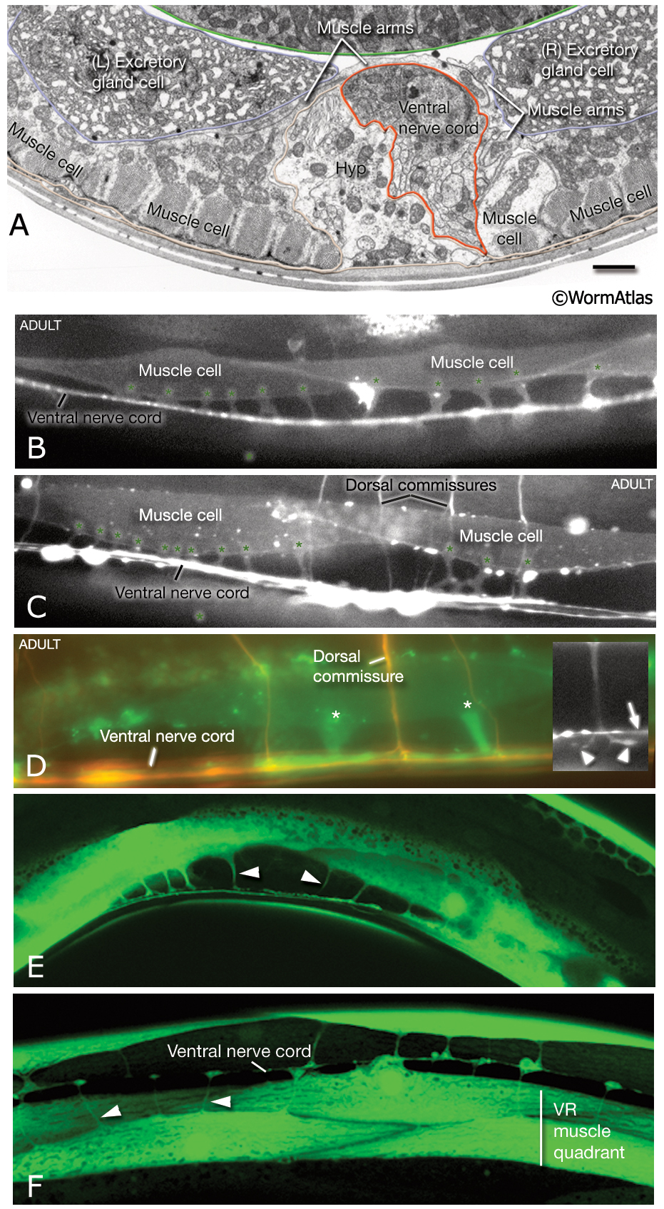
MusFIG 6A-F: Muscle arms of the body wall muscles.
A. Transverse-section TEM from the posterior head region. A muscle arm from the left-side neck muscles is seen crossing over the ventral hypodermal ridge and the ventral nerve cord (red lines) to receive innervation at the right edge of the major fascicle of the ventral nerve cord. Bar, 1 μm. (Image source: N2U [MRC] 240-18.)
B-D. Epifluorescent micrographs from adult transgenic animals co-expressing the him-4p::MB::YFP (muscle), hmr-1b:: DsRed2 (neuron) and unc-129nsp:: DsRed2 (neuron) reporter genes. All are lateral views of the body. Each muscle cell extends 3-6 muscle arms (asterisks in D), mainly from the middle region of each cell. Upon reaching the nerve cord (inset inD, arrow) the muscle arm often bifurcates (inset in D arrowheads) and spreads along the basal lamina to interdigitate and receive input from cord motor neurons. (Strain source: P. Roy.)
E&F. Epifluorescent micrographs from adult transgenic animals expressing the ZK822.5::GFP reporter. E. Lateral view shows slender muscle arms (arrowheads) reaching the ventral nerve cord. F. Ventral view, posterior body. Muscle arms extend from the each of the ventral muscle quadrants (only the right side is labeled) to the ventral nerve cord. The muscle cells that lie in outer rows extend their arms over their partners in the quadrant (arrowheads). (Image source: R. Newbury. The Genome BC C. elegans gene expression consortium [McKay et al, 2004].)
See also MusFIG 6G-K.
Click on picture for full resolution image.
|