|
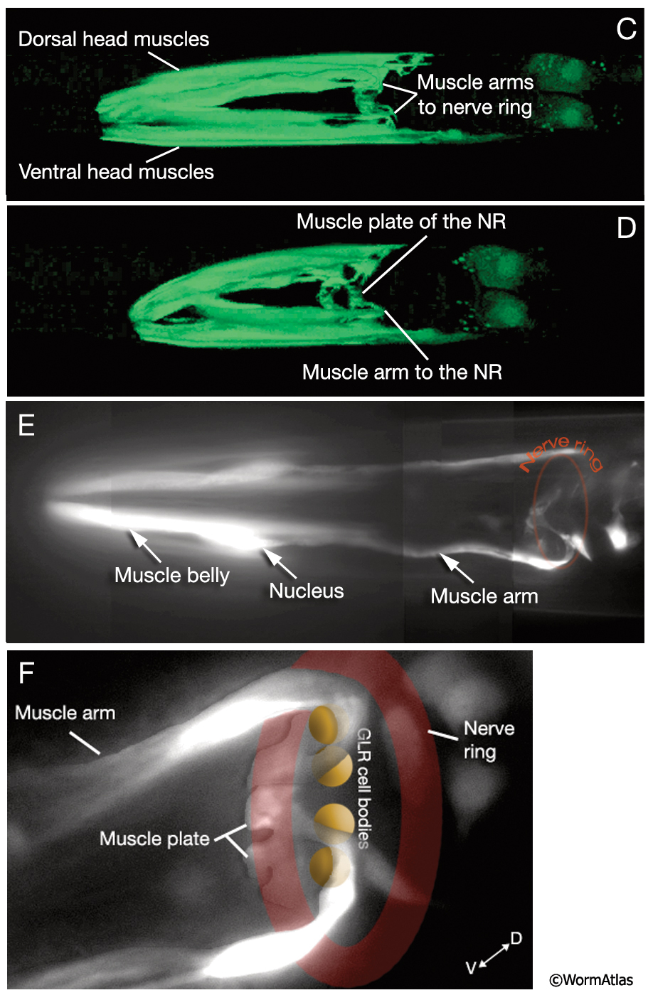
MusFIG 5C-F: Muscle arms of the head muscles.
C & D. Head muscles and muscle arms. Epifluorescent images from a transgenic animal expressing the W05E10.4::GFP reporter gene. C. Left lateral view. D. Left lateral oblique view. The cylindrical muscle plate lying under the nerve ring is clearly seen. (Image source: R. Newbury. The Genome BC C. elegans gene expression consortium [McKay et al, 2004].)
E & F. Anteriormost head muscles, ventral views. Epifluorescent images from a transgenic animal in which GFP is concentrated in nonsarcomeric portions of the muscle cell. E. The muscle arm from a muscle cell in the anterior ventral left quadrant is seen reaching the posterior of the nerve ring region, where it makes a turn to reach inside the nerve ring. F. The same animal, with the nerve ring region is shown in higher magnification. The ventral and lateral GLR cell bodies and the nerve ring are pseudocolored over the epifluorescent image to show their positions relative to the muscle arms and muscle plate. Muscle arms are thought to be guided by the GLR cell bodies to their correct positions along the nerve ring. Original magnification, 600x. (Strain source: M. Land and C. Rubin.) See a 3-D reconstruction of head muscles and muscle arms created by R. Newbury & Moerman lab using Zeiss LSM 5 Pascal software v. 3.2 from confocal images of a strain expressing the GFP marker linked to the promoter for W05E10.4.
See also MusFIG 5A&B and MusFIG 5G.
Click on picture for full resolution image.
|