|
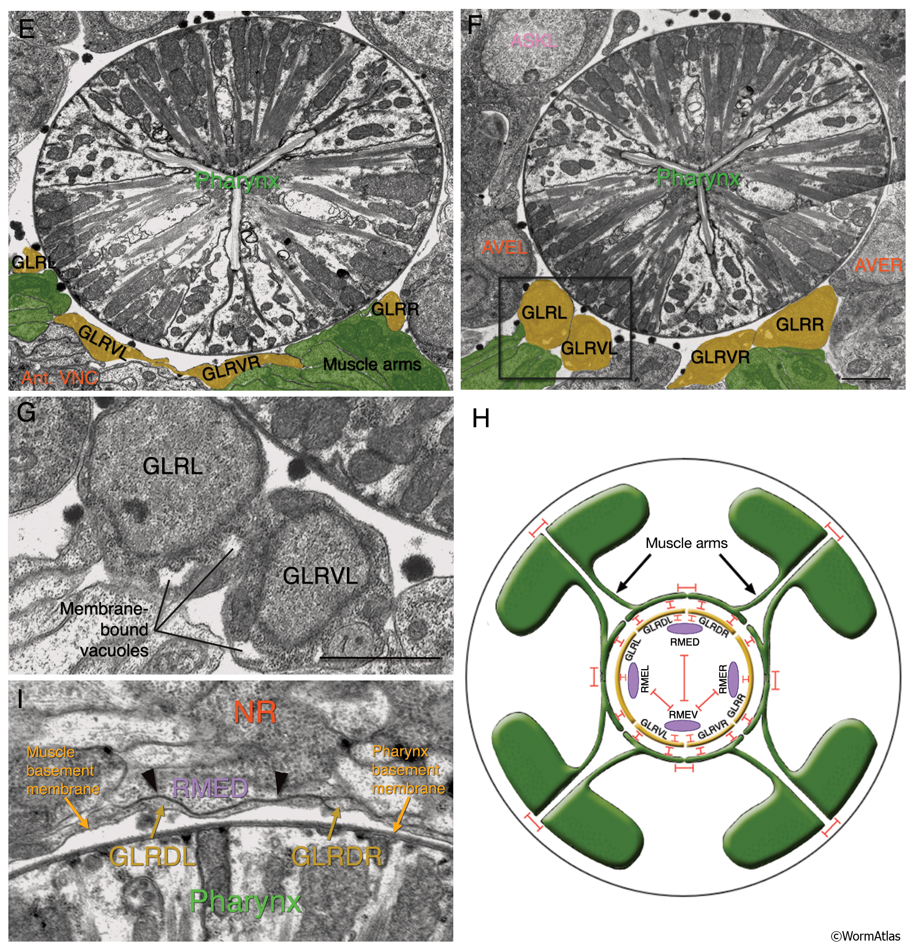
GlrFIG 3E-I: Fine structure of the GLR cells.
All except H are transverse-section TEMs. (NR) Nerve ring. Bars, 1 μm.
E. Because of the anterior tilt of the NR on the dorsal side, the cell bodies of the lateral and ventral GLR cells are placed more posteriorly than those of the dorsal pair. Seen at this level is the broad neck of each lateral and ventral GLR cell connecting the soma to the thin anterior process. (Image source: N2U [MRC] A201 1–4.)
F. At the level of ASK and AVE cell bodies, the cell bodies of the lateral and ventral GLR cells are clustered on the ventral side. (Image source: N2U [MRC] A204 9–12.)
G. Higher magnification of the section in F. Membrane-bound vacuoles are seen in GLRL and GLRVL cell bodies.
H. GLR cells make extensive gap junctions (red bars) to the muscle arms and to RME neurons as shown. For stylistic reasons, RME processes are shown inside the GLR cell layer. In actuality, they lie outside the GLRs and muscle plate. There are also gap junctions between RME neurons and between the muscle bellies of the muscles. No gap junctions are seen between the muscle arms of cells within the same quadrant, but gap junctions exist between arms of cells in different quadrants. (Based on White et al., 1986.)
I. In the anterior regions of the nerve ring where the muscle arm plate is not present anymore, the GLR cell processes make contacts with RME processes and make gap junctions (arrowheads) to them. (Image source: N2U [MRC] A178 10.)
See also GlrFIG 3A-D.
Click on picture for full resolution image.
|