|
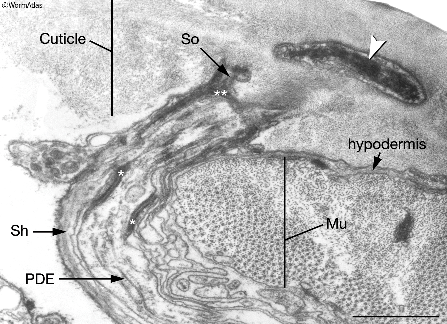
IntroFIG 4B: Transmission electron microscopy (TEM), transverse section of a posterior deirid sensillum.
Arrowhead points to cytoplasmic material (tubule-associated material, TAM) within PDE (posterior deirid) neuron ending in the cuticle. The neuron is concentrically surrounded by the sheath (Sh) and socket (So) cells, which connect to each other by adherens junctions (single asterisks). Socket cells, in turn, are connected to the hypodermis by adherens junctions (double asterisks). Bar: 1 μm. (Image source: [MRC] N2Y 2761-15.)
See also IntroFIG 4A and 4C.
Click on picture for high resolution image. |