|
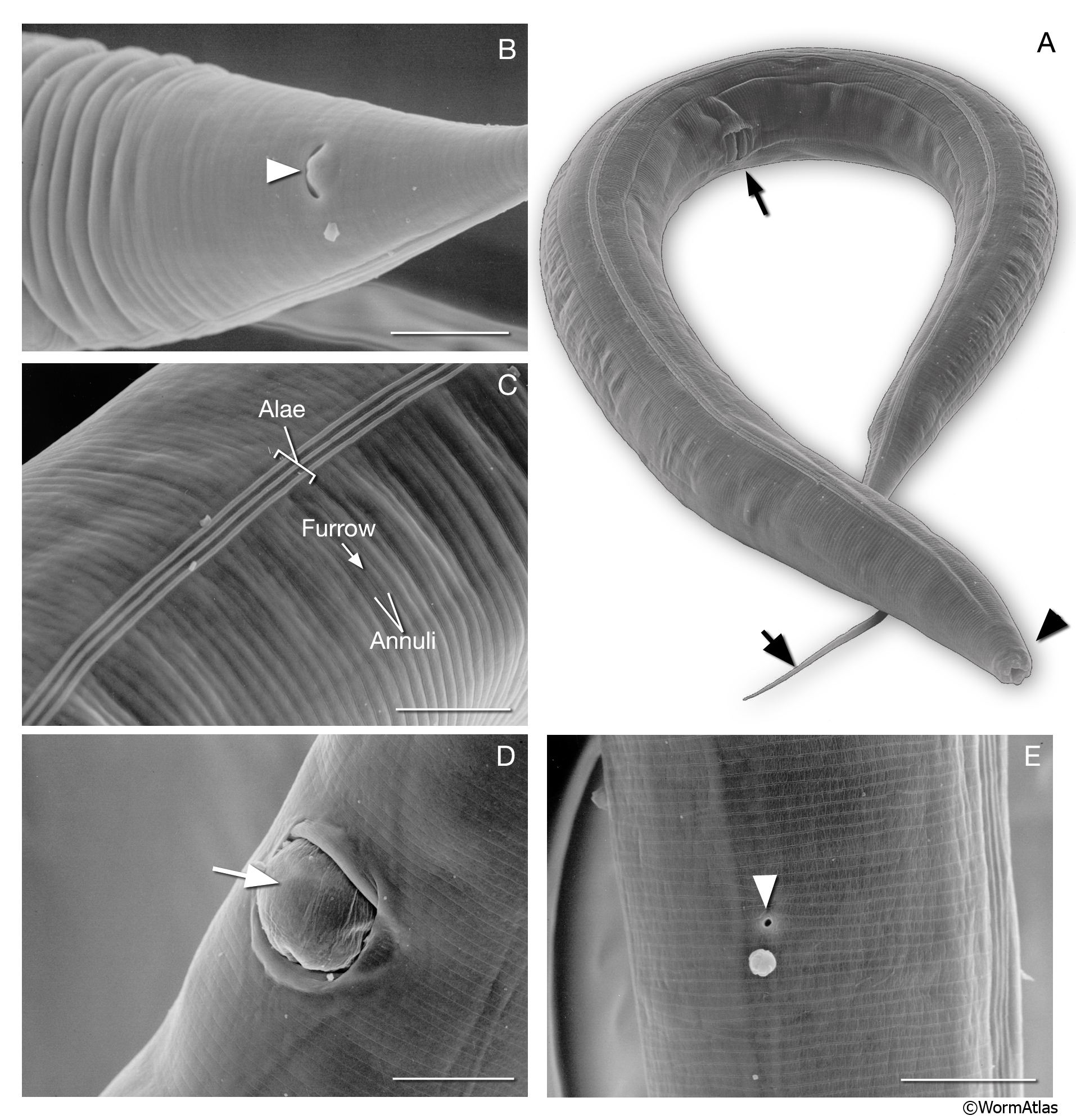
IntroFIG 3: Scanning electron microscopy (SEM) images of adult C. elegans and various body regions.
A. Adult hermaphrodite lying on its right lateral side. (Arrowhead) Lip; (arrow); tail, (thin arrow) the vulva at midbody. (Image reprinted with permission: Juergen Berger and Ralf Sommer, Max Planck-Institute for Developmental Biology, Tübingen, Germany.)
B. Alimentary canal opens to outside through anus at ventral midline (arrowhead). Magnification: 2300x. Scale bar: 10 μm. (Image source: SEM [Hall] 4605.)
C. Outside surface of cuticle on the lateral side bearing circumferential ridges (annuli) and furrows. Alae form over the seam cells. Magnification: 2200x. Scale bar: 10 μm. (Image source: SEM [Hall] 4603.)
D. An egg (arrow) being expelled from vulva. Magnification: 2300x. Scale bar: 10 μm. (Image source: SEM [Hall] 4610.)
E. Excretory pore (arrowhead) located at the ventral midline of head. Magnification: 2700x. Scale bar: 10 μm. (Image source: SEM [Hall] 4609.)
Click on picture for high resolution image. |