|
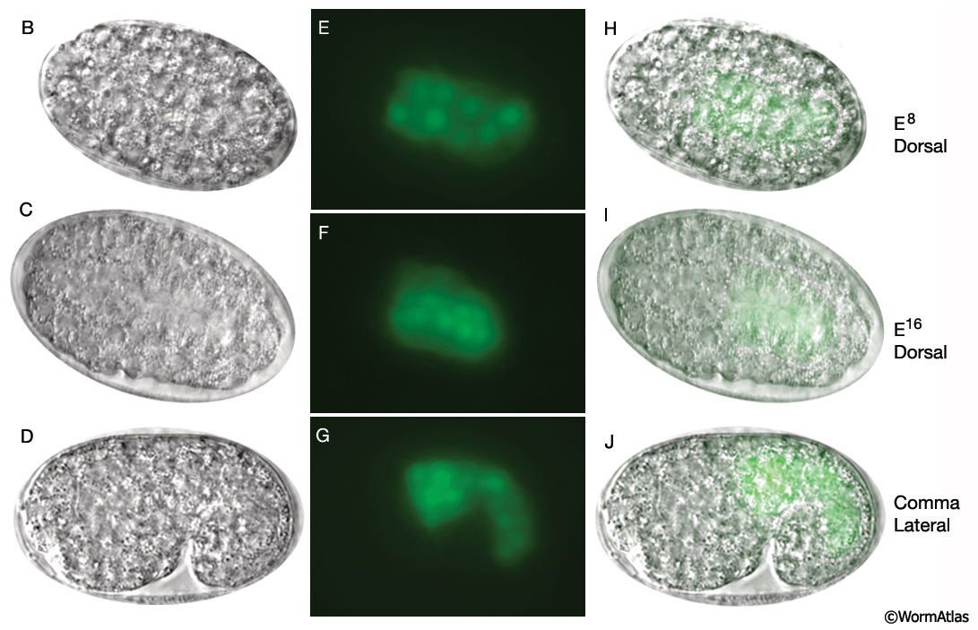
IntFIG 4B-J: Development of the intestine from E8 to L2 stages.
Anterior is to the left. (B-D, K-M, T and V) DIC images. (E-G, N-P, U and W) Epifluorescent images of transgenic animals expressing the reporter gene pW02H5.8-NLS::GFP. (H-J and Q-S) Merged DIC and epifluorescent images. (Pink dots) Basal lamina, detected on the intestinal surface beginning in early morphogenesis. (Strain source: M. Molin, A. Blomberg and M. Pilon.) Magnification, 600x.
See also IntFIG 4A and 4K-W.
Click on picture for full resolution image.
|