|
InterFIG 2: Anterior and posterior arcade cells (as deduced from N2T adult animal).
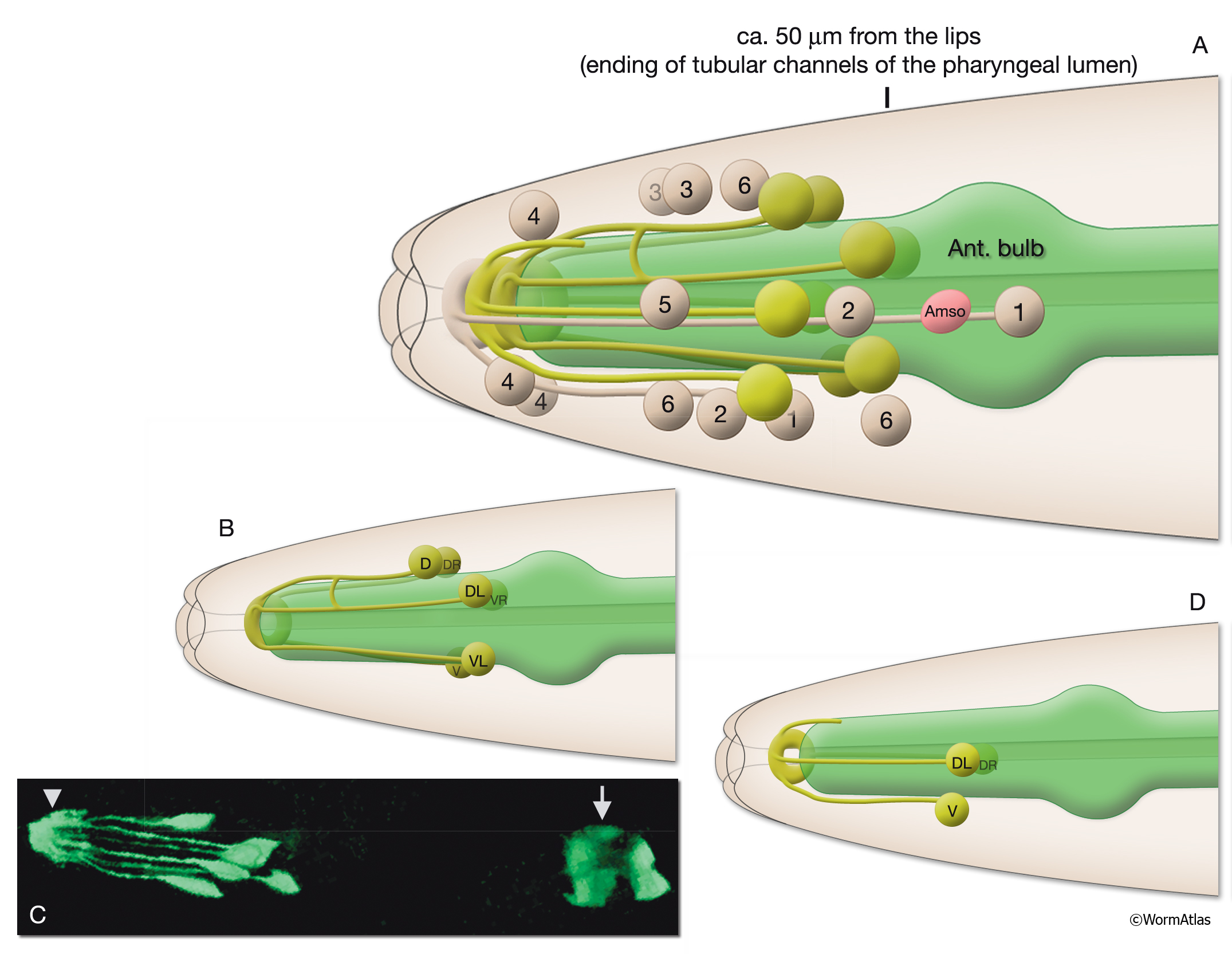
A. The localization of the arcade cells (lime green), the left amphid socket (Amso, pink) and the left lateral, dorsal and ventral midline anterior hypodermal cells (beige; numbered according to the hypodermis type), left lateral view. Anterior and posterior syncytial arcade rings cover the region between pharyngeal epithelium and the hyp 1 epithelial ring, which is made by hyp 1DR (not shown), hyp1DL and hyp1V. The positions of cells are determined from TEM sections of the N2T animal (MRC archive), and there may be slight variations between individual animals. (Ant. Bulb) Anterior bulb of the pharynx.
Shown are only processes of the arcade and hyp 1 cells.
B. Posterior arcade syncytium is made by six cells (arc post D, arc post DL, arc post DR, arc post V, arc post VL and arc post VR). Arcade cells are similar in shape to anterior hypodermal cells with cell bodies in the body wall at the posterior, a syncytial ring at the anterior, and with thin processes connecting the two.
C. Three-dimensional view created from confocal images of a strain expressing the GFP marker linked to the promoter for C08C3.2, left lateral view. All six posterior arcade cells and the posterior arcade ring (arrowhead) are visible. Terminal bulb of the pharynx (arrow) is also expressing the marker gene. (Image source: R. Viverios & D. Moerman.)
D. Anterior arcade syncytium is made by three cells (arc ant DL, arc ant DR and arc ant V). Similar to the posterior arcade, cell bodies of the syncytium are located posterior to the ring, between the tip and the anterior bulb of the pharynx.
Click on picture for full resolution image.
|