|
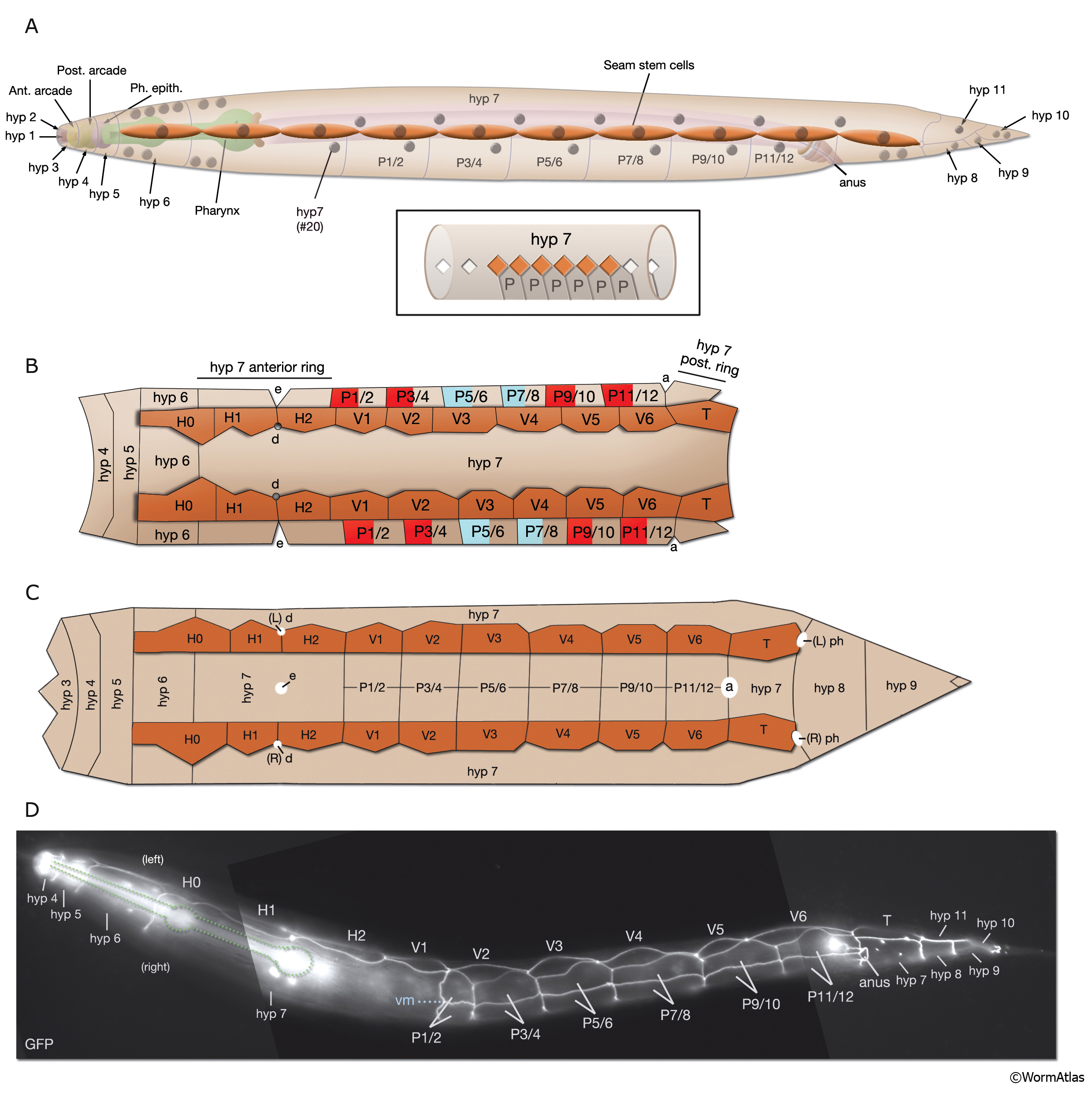
HypFIG 5: Hypodermis in the early L1 stage (until 5 hr post-hatching).
A. Left lateral view of the whole body. At this stage, hyp 7 makes two complete rings wrapping the posterior of the head and the postanal region. Between these two rings, hyp 7 covers only the dorsal and lateral of portions of the body, whereas seam cells and P cells occupy the ventrolateral regions. (Inset) H0, H1, H2, T, hyp 1-6 and tail hypodermis are removed. (Dark circles) Position of nuclei.
B. Animal filleted at the ventral midline. hyp 3 and tail hypodermis (hyp 11, hyp 10) are removed. A single row of ten seam cells is located on each side, extending from the hyp 6-hyp5 junction to the hyp 7-hyp 8 junction. Anterior to hyp 7, hyp 4, hyp 5 and hyp 6 make separate rings of hypodermal tissue. P cells are colored according to the fate of their descendants: P1-P4 and P9-P12 give rise to hypodermis and neurons, whereas P5-P8 generate hypodermis and vulva. (d) Anterior deirid; (e) excretory pore; (a) anus.
C. Animal filleted at the dorsal midline. P1-P12 are aligned in pairs along the ventral midline from anus to the V1-V2 junction. (a) Anus; (ep) excretory pore; (ad) anterior deirid; (ph) phasmid.
D. Epifluorescent image of a transgenic, early L1-stage animal expressing the ajm-1::GFP reporter, ventral oblique view. Visible are apical borders between seam cells, hypodermal cells and P cells as are left side seam cells and P-cell pairs. (Green dotted lines) The pharynx; (vm) ventral midline. Original magnification, 600x. (Strain source: H. Yu and P. W. Sternberg.)
Click on picture for full resolution image.
|