|
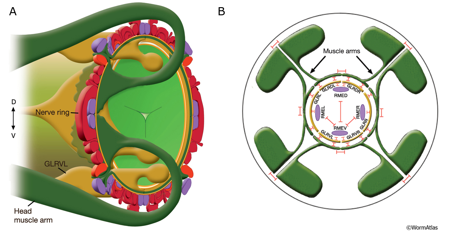
GapjunctFIG 6: Muscle arms of the head muscles are linked by gap junctions.
A. Diagram showing two stylized head muscle arms (dark green) approaching nerve ring. Muscle arms from the 32 muscles in the head and neck project onto the inside surface of the nerve ring in a highly ordered fashion. Their terminal branches lie between the processes of GLR cells (golden yellow) on the inside and the motor neurons of the nerve ring (dark red and purple) on the outside. Arms from the somatic head muscles run posteriorly until they reach the posterior nerve ring region. The arms from each muscle row then make an anterior arc of about 45° and extend inward to reach between the outside surface of the GLRs and the inner surface of the neural plate. This inward turn involves close apposition to the GLR cell bodies. In the neck, somatic muscles extend arms both to the nerve ring and to either the ventral or dorsal nerve cords where they receive additional synapses (not shown). (Light green) pharynx; (orange) basal lamina.
B. GLR cells make extensive gap junctions (red bars) to the muscle arms and to RME neurons as shown. For stylistic reasons, RME processes are shown inside the GLR cell layer. In actuality, they lie outside the GLRs and muscle plate. There are also gap junctions between RME neurons and between the muscle bellies of the muscles, occurring on lateral cell membranes where two muscles meet within the quadrant. No gap junctions are seen between the muscle arms of cells within the same quadrant, but gap junctions exist between arms of cells in different quadrants. (Based on White et al., 1986.)
Click on picture for full resolution image.
|