|
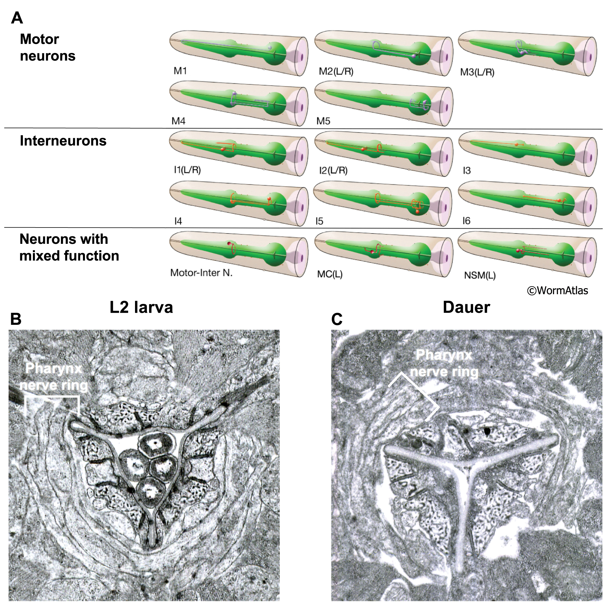
DPhaFIG 10: Neuronal processes within the pharyngeal nerve ring.
A. Illustrations of the pharyngeal neurons. Several pharyngeal neurons extend processes that decussate in the metacorpus to form a pharyngeal nerve ring. (Image source: WormAtlas.)
B&C. The nerve ring is roughly intact in dauers, as compared to a similar region in an L2. There may be a loss of adherence between neuronal and extraneuronal tissues in the dauer, reflected by gaps between the dauer tissues. The pharyngeal pm4 muscles contain dark-staining tubules which are visible around the lumen in L2 and dauer larvae.
B. Transverse TEM section of metacorpus at region of pharyngeal nerve ring in an L2 larva. (Image source: N2 L2 28-14 [D. Riddle] 803.)
C. Transverse TEM section of metacorpus and pharyngeal nerve ring in a dauer larva. (Image source: N2 starved dauer 50-2-1 [D. Riddle] 316M.)
Click on picture for full resolution image.
|