|
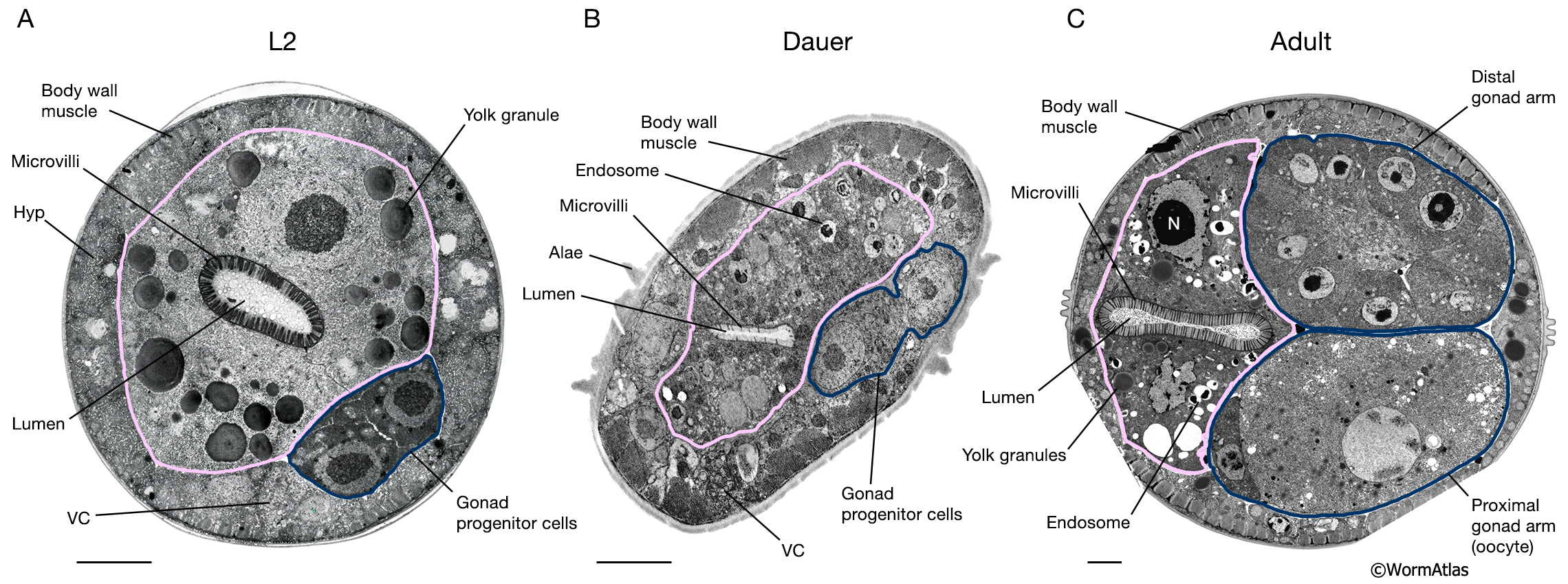
DIntFIG 2A: Midbody intestinal cells in an L2 larva, dauer larva and adult hermaphrodite.
Transverse views of the midbody region in an L2 larva (A), a dauer larva (slightly oblique views) (B) and a mature adult hermaphrodite (C).
A. In larvae, the midbody sections are adjacent to the gonad progenitor cells (outlined in blue).
B.
In the dauer midbody region, the intestinal microvilli are shorter and less dense than in other stages. Intestinal yolk granules appear to be reduced in size and perhaps more electron dense in the dauer. Cytoplasmic endosomes, possibly containing fats or lipids, are also apparent in the dauer intestine.
C. In the adult, components of the mature gonad are visualized (distal and proximal gonad arms, outlined in blue).
Bars,1 micron; VC, ventral nerve cord; Hyp, hypodermis; N, nucleus. (Image sources: A. [N. Thomson] N2 SE1 #191; B. [D. Hall] him-5dar 1127-1 74535 B5 #044; C. [D. Hall] N506 M672.)
Click on picture for full resolution image.
|