|
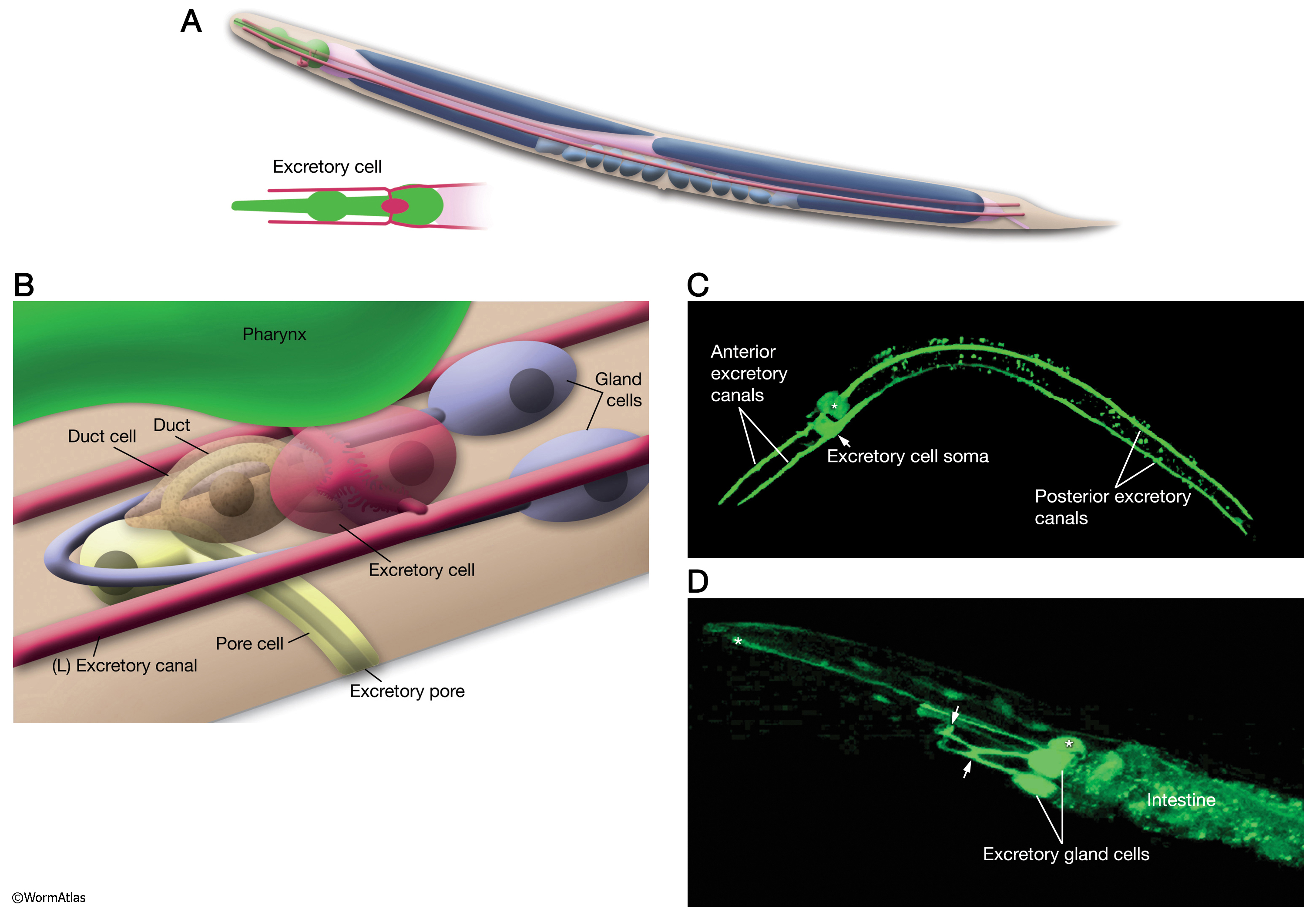
DExcFIG 1: Overview of C. elegans excretory system.
A. Schematic showing the full length of the H-shaped excretory cell as seen from lateral side. In an adult animal, the excretory canals reach from the nose of the nematode to the tail region. (Inset) Head portion from the ventral side.
B. The excretory system (lateral oblique view) consists of the fused pair of gland cells (blue), the excretory (canal) cell (red), the duct cell (brown) and the pore cell (yellow). Somata of all of these cells are located in the head region. The excretory cell is the largest cell in the nematode and is located juxtaposed to the terminal bulb of the pharynx on the ventral side.
C. Epifluorescent image of a transgenic adult animal (dorsal oblique view) expressing the F22E10.1::GFP reporter in the excretory cell. Asterisk marks the pharyngeal gland, which also expresses GFP. (Image source: R. Newbury and D. Moerman.)
D. Epifluorescent micrograph of an animal showing expression of B0403.4::GFP in the excretory gland cell, ventral oblique view. The two small arrows point to regions where two cells fuse to make one syncytial cell. The anterior arrow marks the region where the excretory gland cell is suggested to receive synaptic input from the nerve ring. Pharyngeal gland cells also express this marker (asterisks). (Image source: R. Newbury and D. Moerman.)
Click on picture for full resolution image.
|